
eBook - ePub
Levick's Introduction to Cardiovascular Physiology
Neil Herring, David J. Paterson
This is a test
Buch teilen
- 426 Seiten
- English
- ePUB (handyfreundlich)
- Über iOS und Android verfügbar
eBook - ePub
Levick's Introduction to Cardiovascular Physiology
Neil Herring, David J. Paterson
Angaben zum Buch
Buchvorschau
Inhaltsverzeichnis
Quellenangaben
Über dieses Buch
A sound knowledge of cardiovascular physiology is fundamental to understanding cardiovascular disease, exercise performance and may other aspects of human physiology. Cardiovascular physiology is a major component of all undergraduate courses in physiology, biomedical science and medicine, and this popular introduction to the subject is intended primarily for these students. A key feature of this sixth edition is how state-of-the-art technology is applied to understanding cardiovascular function in health and disease. Thus the text is also well suited to graduate study programmes in medicine and physiological sciences.
Häufig gestellte Fragen
Wie kann ich mein Abo kündigen?
Gehe einfach zum Kontobereich in den Einstellungen und klicke auf „Abo kündigen“ – ganz einfach. Nachdem du gekündigt hast, bleibt deine Mitgliedschaft für den verbleibenden Abozeitraum, den du bereits bezahlt hast, aktiv. Mehr Informationen hier.
(Wie) Kann ich Bücher herunterladen?
Derzeit stehen all unsere auf Mobilgeräte reagierenden ePub-Bücher zum Download über die App zur Verfügung. Die meisten unserer PDFs stehen ebenfalls zum Download bereit; wir arbeiten daran, auch die übrigen PDFs zum Download anzubieten, bei denen dies aktuell noch nicht möglich ist. Weitere Informationen hier.
Welcher Unterschied besteht bei den Preisen zwischen den Aboplänen?
Mit beiden Aboplänen erhältst du vollen Zugang zur Bibliothek und allen Funktionen von Perlego. Die einzigen Unterschiede bestehen im Preis und dem Abozeitraum: Mit dem Jahresabo sparst du auf 12 Monate gerechnet im Vergleich zum Monatsabo rund 30 %.
Was ist Perlego?
Wir sind ein Online-Abodienst für Lehrbücher, bei dem du für weniger als den Preis eines einzelnen Buches pro Monat Zugang zu einer ganzen Online-Bibliothek erhältst. Mit über 1 Million Büchern zu über 1.000 verschiedenen Themen haben wir bestimmt alles, was du brauchst! Weitere Informationen hier.
Unterstützt Perlego Text-zu-Sprache?
Achte auf das Symbol zum Vorlesen in deinem nächsten Buch, um zu sehen, ob du es dir auch anhören kannst. Bei diesem Tool wird dir Text laut vorgelesen, wobei der Text beim Vorlesen auch grafisch hervorgehoben wird. Du kannst das Vorlesen jederzeit anhalten, beschleunigen und verlangsamen. Weitere Informationen hier.
Ist Levick's Introduction to Cardiovascular Physiology als Online-PDF/ePub verfügbar?
Ja, du hast Zugang zu Levick's Introduction to Cardiovascular Physiology von Neil Herring, David J. Paterson im PDF- und/oder ePub-Format sowie zu anderen beliebten Büchern aus Médecine & Théorie, pratique et référence de la médecine. Aus unserem Katalog stehen dir über 1 Million Bücher zur Verfügung.
Information
1 Overview of the cardiovascular system
1.1 Diffusion: its Virtues and Limitations
1.2 Functions of the Cardiovascular System
1.3 The Circulation of Blood
1.4 Cardiac Output and Its Distribution
1.5 Introducing ‘Hydraulics’: Flow, Pressure and Resistance
1.6 Blood Vessel Structure
1.7 Functional Classes of Vessel
1.8 The Plumbing of the Circulation
1.9 Control Systems
• Summary
• Further Reading
LEARNING OBJECTIVES
After reading this chapter you should be able to:
- outline the distance limitation of diffusive transport and the roles of diffusion versus convection in oxygen transport (1.1);
- list the differences between the pulmonary and systemic circulations (1.3);
- sketch out how blood pressure (BP), velocity and total cross-sectional area change from the aorta to the microcirculation and to the vena cava (Figure 1.10);
- write down the basic law of flow (1.5) and apply it to work out the main source of vascular resistance; sketch the structure of the blood vessel wall (Figure 1.11) and state the roles of the endothelium, elastin, collagen and vascular smooth muscle;
- name five main functional categories of blood vessel and state their roles (1.7);
- define a ‘portal circulation’ and explain its functional value (1.8).
The heart and blood vessels evolved to transport O2, nutrients, waste products and heat around the body rapidly. This is crucial for tissue viability, so the cardiovascular system (C VS) develops at an early stage in the embryo. However, very tiny organisms lack a circulatory system - their O2 needs are satisfied by diffusion from the environment. Even large animals, such as humans, rely on diffusion for the transport of materials between the bloodstream and cells. Why, then, do we also need a CVS? The answer lies in the distance limitation of diffusive transport.
1.1DIFFUSION: ITS VIRTUES AND LIMITATIONS
Diffusion is brought about by a molecular ‘drunkard’s walk’
Diffusion is a ‘passive’ transport process, in the sense that it is driven by the rapid, random thermal motion of molecules, not by metabolic pumps. When a concentration gradient is present, the randomly directed step movements of individual solute molecules result in a net movement down the concentration gradient, i.e. a net diffusive transport (Figure 1.1).
Distance dramatically slows diffusion
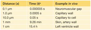
The rate of diffusive transport is important because nutrient delivery must keep up with cellular demand. Fortunately, diffusive transport is very fast over short distances; for example, diffusion from a capillary to tissue cell, a distance of ~10 p m, takes only ~50 ms. Unfortunately, as Einstein showed, the time t that randomly jumping particles take to move a distance x, in one specific direction, increases as the square of the distance:
(1.1)
Thus, diffusion is incredibly slow over long distances (Table 1.1). Over 1 cm, which is the thickness of the human left ventricle wall, diffusion would take more than half a day. Sadly, nature often reminds us that Einstein’s equation is correct. Figure 1.2 shows a section through a human heart after a coronary artery thrombus (clot) had blocked off the blood supply to the left ventricle wall. The pale area is cardiac muscle that died from lack of O2, even though the adjacent chamber is full of oxygenated blood. The patient died because just a few millimetres reduced the rate of diffusive O2 transport to a level that was too low to support life.
Table 1.1 Time taken for a glucose molecule to diffuse specified distance in one direction
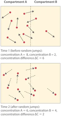
Figure 1.1 Spontaneous molecular steps in a random direction lead to a net movement of solute molecules (dots) down a concentration gradient. The probability of a randomly directed step from compartment A to B is greater than from B to A because there are more solute molecules in A than B, per unit volume. Note that an individual molecule, such as the top one in B, may move ‘uphill’, that is, into the more concentrated solution. Net diffusion is thus the result of unequal ‘uphill’ and ‘downhill’ fluxes.
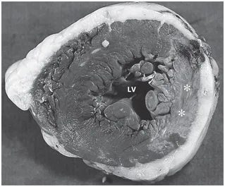
Figure 1.2 Section of the human left ventricle after a coronary thrombosis; the myocardium has been stained for a muscle enzyme. The pale area (marked with two *) is an ‘infarct’, an area of muscle damaged or killed by lack of O2. The pallor is due to the escape of enzymes from the dying muscle. The infarct was caused by a coronary artery obstruction, which halted the convective delivery of O2. O2 diffusion from blood in the main chamber (LV) is unaffected, yet only a thin rim of adjacent tissue (~1 mm) survived. (Courtesy of the late Professor M Davies, St George’s Hospital Medical School, London.)
Source: Einstein A. Investigations on the Theory of the Brownian Movement (trans. by Furth R, Cowper AD, 1956). New York: Dover Publications; 1905.
a Einstein’s equation states t = x 2/2D, where D is solute diffusion coefficient (glucose, 0.9 x 10⁻⁵ cm2 s−1 at 37° C; oxygen in water, 3 x 10⁻⁵ cm2 s−1, 37° C).
Convection provides fast transport over long distances
For distances of >0.1 mm, a faster transport system is clearly needed. The CVS provides this (Figure 1.3). The CVS still relies on diffusion to transport O2 across the short distance between gas and blood in the lungs; however, the absorbed O2 is then washed rapidly along in a stream of pumped fluid, covering a large distance in seconds (~3 cm s−1). This form of transport is called bulk flow or convective transport, and its energ y source is the contraction of the heart. Convective transport carries O2 a metre or more from the lungs to the smallest blood vessels of the human extremities in ~30 s, whereas diffusion would take more than 5 years! Nevertheless, diffusion takes over as the dominant transport process for the final 10-20 pm from blood to cell.
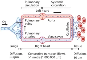
Figure 1.3 Overview of the human circulation, highlighting the relative roles of diffusion and convection in O2 transport.
1.2FUNCTIONS OF THE CARDIOVASCULAR SYSTEM
There are four main functions of the cardiovascular system, as detailed below:
- The primary function of the CVS is the rapid convective transport of O2, glucose, amino acids, fatty acids, vitamins, drugs and water to the tissues, and the rapid washout of metabolic waste products from the tissues (e.g. carbon dioxide (CO2), urea, creatinine).
- The CVS is also a control system. It distributes hormones to the tissues and secretes bioactive agents itself (natriuretic peptides, renin, nitric oxide, endothelin, prostaglandins).
- The CVS is crucial for body temperature regulation because it transports heat from deep organs to the skin surface and regulates heat loss from the skin.
- In reproduction, the CVS provides the hydraulic mechanism for genital erection.
1.3THE CIRCULATION OF BLOOD
The heart consists of two synchronous, muscular pumps, the right and left ventricles (Figure 1.4). Each pump is filled from a contractile reservoir, the right or left atrium. The right ventricle pumps deoxygenated blood through the pulmonary trunk to the lungs (Figure 1.5). Four pulmonary veins return oxygenated blood from the lungs to the left side of the heart, completing the short, low-pressure pulmonary circulation. The left ventricle pumps an equal volume of oxygenated blood to the tissues of the body. The tissues extract some of the O2, and the partly deoxygenated blood returns via two great veins, the superior and inferior vena cava, to the right atrium. This completes the long, high-pressure systemic circulation. One-way valves in the he...