Gregory Y. Lauwers, Michael B. Wallace, Gregory Y. Lauwers, Michael B. Wallace
This is a test
This is a test
Buch teilen
English
ePUB (handyfreundlich)
Über iOS und Android verfügbar
eBook - ePub
Gastrointestinal Pathology
Correlative Endoscopic and Histologic Assessment
Gregory Y. Lauwers, Michael B. Wallace, Gregory Y. Lauwers, Michael B. Wallace
Angaben zum Buch
Buchvorschau
Inhaltsverzeichnis
Quellenangaben
Über dieses Buch
An illustrated guide to best practices when performing and assessing biopsies for GI conditions of all kinds
Accurate diagnosis of GI conditions necessarily entails both the careful taking of biopsies and the informed analysis of tissue material. With that being so, gastroenterologists and GI pathologists alike must have a solid understanding of the techniques, handling requirements, and diagnostic characteristics involved if they are to collaborate effectively. Gastrointestinal Pathology has been designed to provide a clinically focussed and richly illustrated guide to real-world scenarios faced by practicing GI specialists, offering step-by-step instruction and professional advice on the correct diagnosis of all major GI conditions. This essential new book includes:
Full-color illustrations throughout
Complete details of biopsy samples required to diagnose specific conditions
Reviews of differential diagnoses
Clinical management clues based on pathologic findings
Featuring information to improve the practice of all gastroenterologists and GI pathologists, Gastrointestinal Pathology is a practical and every-day resource for the precise diagnosis of a wide range of GI conditions.
Häufig gestellte Fragen
Wie kann ich mein Abo kündigen?
Gehe einfach zum Kontobereich in den Einstellungen und klicke auf „Abo kündigen“ – ganz einfach. Nachdem du gekündigt hast, bleibt deine Mitgliedschaft für den verbleibenden Abozeitraum, den du bereits bezahlt hast, aktiv. Mehr Informationen hier.
(Wie) Kann ich Bücher herunterladen?
Derzeit stehen all unsere auf Mobilgeräte reagierenden ePub-Bücher zum Download über die App zur Verfügung. Die meisten unserer PDFs stehen ebenfalls zum Download bereit; wir arbeiten daran, auch die übrigen PDFs zum Download anzubieten, bei denen dies aktuell noch nicht möglich ist. Weitere Informationen hier.
Welcher Unterschied besteht bei den Preisen zwischen den Aboplänen?
Mit beiden Aboplänen erhältst du vollen Zugang zur Bibliothek und allen Funktionen von Perlego. Die einzigen Unterschiede bestehen im Preis und dem Abozeitraum: Mit dem Jahresabo sparst du auf 12 Monate gerechnet im Vergleich zum Monatsabo rund 30 %.
Was ist Perlego?
Wir sind ein Online-Abodienst für Lehrbücher, bei dem du für weniger als den Preis eines einzelnen Buches pro Monat Zugang zu einer ganzen Online-Bibliothek erhältst. Mit über 1 Million Büchern zu über 1.000 verschiedenen Themen haben wir bestimmt alles, was du brauchst! Weitere Informationen hier.
Unterstützt Perlego Text-zu-Sprache?
Achte auf das Symbol zum Vorlesen in deinem nächsten Buch, um zu sehen, ob du es dir auch anhören kannst. Bei diesem Tool wird dir Text laut vorgelesen, wobei der Text beim Vorlesen auch grafisch hervorgehoben wird. Du kannst das Vorlesen jederzeit anhalten, beschleunigen und verlangsamen. Weitere Informationen hier.
Ist Gastrointestinal Pathology als Online-PDF/ePub verfügbar?
Ja, du hast Zugang zu Gastrointestinal Pathology von Gregory Y. Lauwers, Michael B. Wallace, Gregory Y. Lauwers, Michael B. Wallace im PDF- und/oder ePub-Format sowie zu anderen beliebten Büchern aus Medicine & Gastroenterology & Hepatology. Aus unserem Katalog stehen dir über 1 Million Bücher zur Verfügung.
1 General Principles of Biopsy Diagnosis of GI Disorders
Herbert C. Wolfen1, Michael B. Wallace1, Naohisa Yahaghi2 and Yutaka Saito3
1 Mayo Clinic, Jacksonville, Florida, USA
2 Keio University Cancer Center, Tokyo, Japan
3 National Cancer Center, Tokyo Japan
Tissue sampling of the gastrointestinal tract at the time of endoscopy is the cornerstone of many gastrointestinal diagnoses. The development of a flexible endoscope and the subsequent ability to directly acquire tissue under optical guidance has been one of the most important advancements in the field of gastroenterology throughout its history. Although tissue sampling can be performed through nonendoscopic devices, the ability to directly correlate precise locations and target biopsies to specific areas of disease is critical to our ability to diagnose and further understand gastrointestinal pathology. Many of the advancements in our understanding of the basic pathology and molecular biology of gastrointestinal disease can be directly attributed to our ability to acquire tissue for histological, molecular, and genetic analyses. An excellent example is our deep understanding of the molecular pathology of colorectal cancer development from normal colonic epithelium to adenoma to colorectal cancer, a discovery made possible because of colonoscopic access to precursor lesions such as adenomatous polyps and early cancers.
In this chapter, we will review general principles of tissue acquisition at the time of endoscopy including the following topics:
Endoscopic equipment for obtaining tissue including endoscopic accessory channels, biopsy forceps, snare devices, needle aspiration and cytology brush.
General principles of optimal sampling technique.
Methods of tissue preparation in the endoscopy laboratory to optimize diagnostic accuracy.
The role of endoscopic ultrasound (EUS)‐guided fine‐needle aspiration cytology.
Endoscopic Equipment for Tissue Sampling
Modern endoscopic equipment can be divided in two general categories: the endoscope that allows access to the gastrointestinal tract and accessory devices that are typically passed through the working channel of the endoscope to directly acquire tissue, including biopsy forceps, snares, fine‐needle aspiration devices, and cytology brushes. Recent developments in tissue sampling include devices that are capable of wide‐field, often definitive, endoscopic resection of early neoplasia and invasive carcinoma.
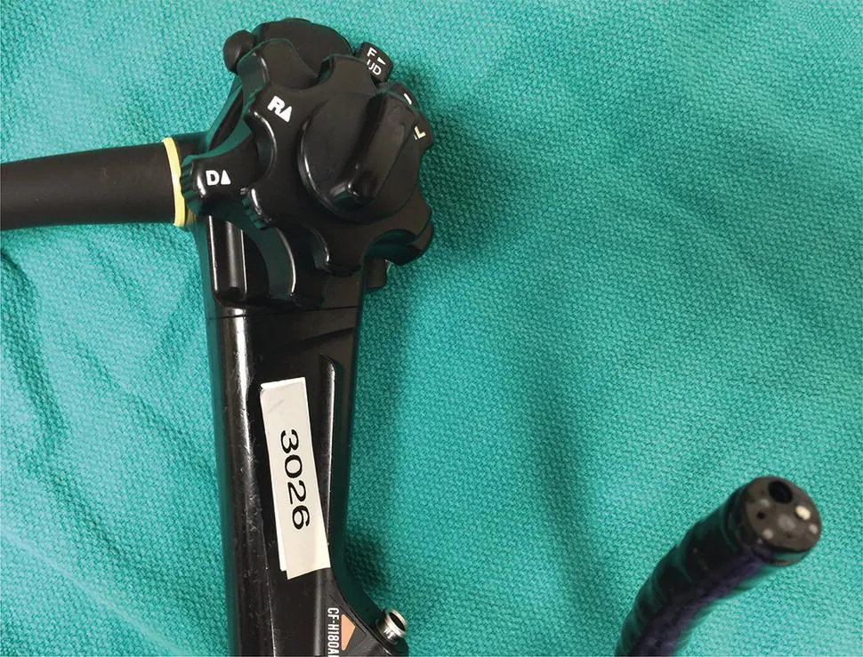
A modern endoscope is a remarkably robust and versatile instrument including a light source, optical lenses with a video capture device, image processing, and display equipment, and importantly for the purposes of tissue acquisition, an accessory channel ranging from 1 to 6 mm (typically 3–4 mm), which allows passage of devices for mechanical collection of tissue (Figures 1.1 and 1.2).
There is a general trade‐off between the diameter of the instrument and the ease and comfort with which it can be passed through the natural orifices of the body such as the mouth and anus. In general the larger the outer diameter, the larger the accessory channel is to accommodate larger instruments for tissue acquisition. A fundamental limitation of most flexible endoscopes, as opposed to surgical instruments, is that all accessories pass through a single access point of the endoscope. As compared to surgical instruments with multiple access points, the endoscopic devices do not typically allow triangulation to acquire a large bulk tissue or resect entire organs. For this reason, most tissue is sampled through pinch forceps, needle aspiration, or wire loop snare devices. More recently, electrosurgical needles and other cutting tools have been developed, which have allowed wide‐field resection of tissues of virtually any diameter (Figure 1.3).
Figure 1.1 Endoscope with control handle and tip. The tip contains a light source, imaging window, and accessory channel through which various tissue acquisition devices can be passed.
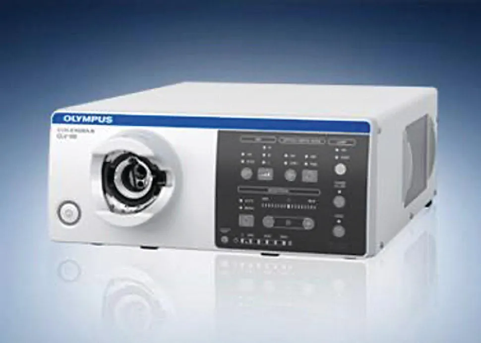
Figure 1.2 Endoscopic processor, which converts the light captured from the endoscope tip into a visible image for display.
Source: Olympus America, Inc. With permission.
Figure 1.3 (a) Tools for performing endoscopic resection including endoscopic submucosal dissection (ESD).
Source: Zeon Medical.
(b) Standard and insulated tip electrocautery knives for incision and dissection.
(c) CO2 insufflator for luminal distension, which is preferred to air given rapid reabsorption.
Source: Olympus.
(d) Distal attachment hood to facilitate maintaining view within the submucosal space.
Source: Fujifilm medical.
(e) Injection fluid (hyaluronic acid; Mucoup [Johnson and Johnson]) for submucosal lifting.
Source: Gut and Liver.
Pinch Biopsy Forceps
The flexible pinch biopsy forceps have been one of the most versatile of all instruments for tissue acquisition. These typically involve a flexible steel cable and lever device with two sharp‐edged cups, which can be opened and closed to acquire tissue (Figure 1.4).
Standard endoscopic sampling typically acquires tissue from the mucosa and occasionally a submucosal depth of the intestinal wall; however, large‐capacity forceps as well as multiple sampling including “bite on bite” allow sampli...