Type 1 Reaction (T1R)
Cleverson Teixeira Soares Abstract
A type 1 reaction (T1R) is also known as a reversal reaction. This phenomenon involves exacerbation of the immune system or delayed-type hypersensitivity in response to the antigens of Mycobacterium leprae present in parasitized tissues. It occurs in most patients of the tuberculoid and borderline forms of the Ridley & Jopling classification for leprosy. It is an important phenomenon that can occur before, during, or after leprosy treatment and can be destructive, causing tissue damage mainly in the nerves, as well as irreversible sequelae. The recognition of T1R in histological sections may be notified prior to clinical presentation. Histopathological recognition is vital in defining or confirming the presence of T1R, guiding the treatment of the reaction process, avoiding or reducing the possibility of serious sequelae, correcting possible mistakes in the classification of patients within the spectrum of leprosy, and ruling out other diseases that can clinically simulate a T1R. In this chapter, the histopathological characteristics that allow the recognition of T1R, various histopathological aspects of the common forms of leprosy, and histopathological differential diagnoses are discussed.
Keywords: Downgrading, Hansen’s disease, Leprosy, Reaction type 1, Reversal reaction, Upgrading.
INTRODUCTION
Leprosy is a slow and progressive disease noted by inflammatory signs on the skin as lesions and in the peripheral nervous system. Granulomas develop slowly, allowing endoneural structures and other parasitic tissues to adapt to the immune response. Functional changes are noticed after long-term disease progression as a result of the low antigenicity of M. leprae that limits an acute, intense, or destructive immunocelluar reaction. However, highly intense and destructive episodes involving the abrupt onset of cutaneous-neural lesions may appear during the course of the disease. These episodes are called leprosy reactions.
Reactions are important events that occur throughout the progression or regression of leprosy. To date, there is no specific treatment to prevent the occurrence of these epiphenomena, nor an effective treatment protocol for all cases [1-3]. Generally, during these episodes, neurological lesions can worsen
and cause permanent functional disabilities [1, 2]. There are two main types of reactions identified in leprosy: type 1 reaction (T1R) and type 2 reaction (T2R). In this chapter, the histopathological characteristics of T1R will be discussed.
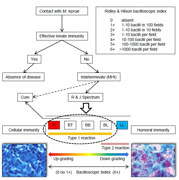
Fig. (1)) Clinical spectrum and bacilloscopic index of leprosy forms and reactions. Patients who are exposed to
M. leprae can eliminate the bacilli through mechanisms of primary immune response and do not develop the disease. If the primary immune defense cannot contain the proliferation of the bacilli, the patient develops indeterminate leprosy (I), the early stage of the disease preceding the polarized forms of the Ridley & Jopling (R&J) classification: tuberculoid (TT), borderline-tuberculoid (BT), borderline-borderline (BB), borderline-lepromatous (BL) and lepromatous or virchowian (LL). Late recognition of bacillary antigens by the individual may result in an intense and effective immune response (TT and BT pattern), which may lead to the destruction of the bacilli and spontaneous cure. TT individuals are those with effective cellular immunity. If cellular immunity is not effective, proliferation and dissemination of the bacilli persist, and the disease progresses toward the lepromatous pole. LL individuals are anergic and react to the bacilli through humoral immunity. Type 1 reactions (T1R) affect patients in the range from TT to BL. Type 2 reactions (T2R) affect patients on the lepromatous side (BL and LL). The bacilloscopic index ranges from 0-6+.
Histopathological and Bacilloscopic Characteristics of T1R
T1R is an exacerbation of the immunocellular response that occurs in patients in the TT, BT, BB, and BL forms (Fig. 1). The clinical signs of the reaction are swelling and erythema over pre-existing lesions (Fig. 2). Some skin lesions that are difficult to identify or clinically imperceptible may become evident during the reaction process (Fig 2). T1R can occur at different locations on the skin and other parasitic tissues with varying intensity (Fig. 3). Necrosis and ulceration of lesions are characteristics of intense T1Rs, especially when it occurs in patients in the tuberculoid side (TT and BT) (Figs. 4-5 ) [4]. T1Rs are represented histologically by a tuberculoid granuloma, similar to those observed in TT and BT granulomas (“TT/BT-like granuloma”), consisting mainly of M1-pattern epithelioid macrophages, permeated by T lymphocytes and followed by M2-pattern macrophages, B and T lymphocytes and other cells in the periphery (Fig. 6-7) [5]. Macrophage fusion forming multinucleated giant cells is common. Granulomas are confluent and have inaccurate limits. Lymphocytes permeate epithelioid macrophages, which have intracytoplasmic vacuoles and intercellular edema. There may be deposition of interstitial fibrin, focal or confluent necrosis, and various degrees of aggression to the epidermis, with associated epithelial hyperplasia (Fig. 6). These changes are due to the influx of new cells (macrophages, lymphocytes, plasma cells, and other cells) associated with changes that occur in existing cells, which constitutes the inflammatory process of pre-existing lesion leprosy [5]. Therefore, the histopathological and bacilloscopic characteristics of a histological section containing a type 1 reaction lesion are the sum of the histopathological characteristics of the lesion present before the reaction episode (TT, BT, BB, and BL) with the overlap of T1R histopathological characteristics.
Since T1R is an immunocellular reaction with an outline of tuberculoid granulomas, some cases of borderline leprosy with associated T1R can be confused with tuberculoid leprosy (TT). Although similar, the tuberculoid granulomas of T1R can be differentiated from those of the TT and BT forms. Some histological features are present in T1R tuberculoid granulomas and rare or absent in TT/BT tuberculoid granulomas: (1) granulomas are of imprecise and confluent limits; (2) they do not have a lymphocyte mantle on the periphery, especially those located in the reticular dermis or adipose tissue; (3) there are several lymphocytes permeating the epithelioid macrophages in the center of the granulomas; (4) there are important intercellular and intracellular edema in the macrophages in the center granulomas and in the interstitium and (5) fibrinoid or caseous necrosis is present in the center of the granulomas (Figs. 6 , 8-9 ). The erroneous classification of a borderline patient with T1R as TT has important implications for the choice of appropriate treatment, since borderline with T1R patients follow a treatment protocol for multibacillaries and TT patients are treated for paucibacillaries (Figs. 10-11) [6, 7]. If the histological sections show disorganized and confluent tuberculoid granulomas, significant edema, foci of necrosis within the granulomas, extension of the inflammatory process to the interstitium associated with stellated fibroblasts with evident nucleoli and a bacilloscopic index ≥ 2+, this probably represents a T1R over a borderline lesion instead of leprosy in the tuberculoid side (TT/BT). The histopathological characteristics of tuberculoid granulomas of the TT and BT forms involving different skin tissues are detailed in chapters 3 an...