Insect Physiology and Biochemistry
eBook - ePub

Insect Physiology and Biochemistry

James L. Nation, Sr.

Buch teilen
  1. 564 Seiten
  2. English
  3. ePUB (handyfreundlich)
  4. Über iOS und Android verfügbar
eBook - ePub

Insect Physiology and Biochemistry

James L. Nation, Sr.

Angaben zum Buch
Buchvorschau
Inhaltsverzeichnis
Quellenangaben

Über dieses Buch

Employing the clear, student-friendly style that made previous editions so popular, Insect Physiology and Biochemistry, Fourth Edition presents an engaging and authoritative guide to the latest findings in the dynamic field of insect physiology. The book supplies a comprehensive picture of the current state of the function, development, and reproduction of insects. Expanded and updated, now in full colour, this fourth edition adds three new chapters on the role of the nervous system in behavior; the 'Genomics Revolution' in entomology; and global climate changes which have a major effect on insects, including warming and weather. It continues to challenge conventional entomological wisdom with the latest research and analytical interpretations.

The text will appeal to upper undergraduate and graduate students and to practicing biologists who need to possess a firm knowledge of the broad principles of insect physiology. With detailed full colour illustrations to help explain physiological concepts and important anatomical details, it remains the most easily accessible guide to key concepts in the field.

Häufig gestellte Fragen

Wie kann ich mein Abo kündigen?
Gehe einfach zum Kontobereich in den Einstellungen und klicke auf „Abo kündigen“ – ganz einfach. Nachdem du gekündigt hast, bleibt deine Mitgliedschaft für den verbleibenden Abozeitraum, den du bereits bezahlt hast, aktiv. Mehr Informationen hier.
(Wie) Kann ich Bücher herunterladen?
Derzeit stehen all unsere auf Mobilgeräte reagierenden ePub-Bücher zum Download über die App zur Verfügung. Die meisten unserer PDFs stehen ebenfalls zum Download bereit; wir arbeiten daran, auch die übrigen PDFs zum Download anzubieten, bei denen dies aktuell noch nicht möglich ist. Weitere Informationen hier.
Welcher Unterschied besteht bei den Preisen zwischen den Aboplänen?
Mit beiden Aboplänen erhältst du vollen Zugang zur Bibliothek und allen Funktionen von Perlego. Die einzigen Unterschiede bestehen im Preis und dem Abozeitraum: Mit dem Jahresabo sparst du auf 12 Monate gerechnet im Vergleich zum Monatsabo rund 30 %.
Was ist Perlego?
Wir sind ein Online-Abodienst für Lehrbücher, bei dem du für weniger als den Preis eines einzelnen Buches pro Monat Zugang zu einer ganzen Online-Bibliothek erhältst. Mit über 1 Million Büchern zu über 1.000 verschiedenen Themen haben wir bestimmt alles, was du brauchst! Weitere Informationen hier.
Unterstützt Perlego Text-zu-Sprache?
Achte auf das Symbol zum Vorlesen in deinem nächsten Buch, um zu sehen, ob du es dir auch anhören kannst. Bei diesem Tool wird dir Text laut vorgelesen, wobei der Text beim Vorlesen auch grafisch hervorgehoben wird. Du kannst das Vorlesen jederzeit anhalten, beschleunigen und verlangsamen. Weitere Informationen hier.
Ist Insect Physiology and Biochemistry als Online-PDF/ePub verfügbar?
Ja, du hast Zugang zu Insect Physiology and Biochemistry von James L. Nation, Sr. im PDF- und/oder ePub-Format sowie zu anderen beliebten Büchern aus Sciences biologiques & Entomologie. Aus unserem Katalog stehen dir über 1 Million Bücher zur Verfügung.

Information

Verlag
CRC Press
Jahr
2022
ISBN
9781000577457

1 Embryogenesis

DOI: 10.1201/9781003279822-1
Highlights
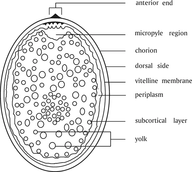
  • A proteinaceous chorion is put on the egg while it is in the ovary.
  • Sperm released from the spermatheca of the female enter the egg through the micropyle, a narrow channel through the chorion, as the egg passes down the oviduct.
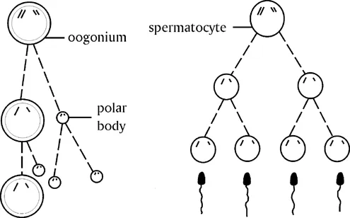
  • Usually, the egg nucleus is diploid until the entry of the sperm stimulates meiotic division leading to the haploid egg nucleus. Union of a sperm nucleus with the egg nucleus produces the zygote and stimulates the zygote to begin divisions.
  • Zygotic divisions result in large numbers of nuclei surrounded with a small field of cytoplasm but without cell membranes; these nuclei and cytoplasm migrate into a single layer near the periphery of the egg and form the blastoderm.
  • Maternal genes are present and active in the nurse cells of the mother during oogenesis, and they pass maternal gene transcripts (mRNAs, tRNAs, proteins) into the developing oocyte in the ovary.
  • Gap genes divide the embryo into large domains; then pair-rule genes divide the domains into parasegments, and finally, segment polarity genes control formation of true segments.
  • Homeotic genes function during parasegment formation and give each segment its characteristic identity.
  • Drosophila is a model for genetic studies of embryogenesis and offers important insights into the development of higher organisms including humans.

1.1 Introduction

The three major divisions in the Insecta, the Apterygota, Hemimetabola, and Holometabola, are not directly ancestral to each other, and consequently embryological developments in the groups, although similar in some respects, often diverge. The Apterygota (Protura, Collembola, Diplura, and Thysanura) never evolved wings and lack metamorphosis. The immatures look like small versions of the adults. The Hemimetabola (Orthoptera, Hemiptera, and others) evolved wings and have gradual metamorphosis, but no pupal stage. Immatures have some adult features but lack fully developed wings until the adult stage. The Holometabola (Coleoptera, Hymenoptera, Lepidoptera, Diptera, and others) evolved adult wings and have complete metamorphosis, with a pupal stage. Immature forms are typically wormlike, and thus look very different from the adults. Wingless adults occur in the Hemimetabola and Holometabola, but the wingless condition evolved secondarily from winged forms.
The goals for this chapter are to describe the morphogenetic events and the action of genes in formation of the embryo. The work by Johannsen and Butt (1941) is still a very valuable source for understanding variations in morphogenesis, as are reviews by Andersen (1972a, 1972b), Jura (1972), Sander et al. (1985), Campos-Ortega and Hartenstein (1985), Panfilio et al. (2006), and Panfilio (2008). A review of morphology of embryogenesis in the silkworm, Bombyx mori, has been published (Miya, 1985), and the early stages of embryogenesis are described for several species of fireflies by Kobayashi and Ando (1985).
The complete analysis of the genome of Drosophila melanogaster (Adams et al., 2000), its many mutants, ease and economy of rearing, and a great host of workers means that more details of the genetics of embryogenesis are available for D. melanogaster than for any other insect, although the genomes of numerous insects are now available. A timeline for some of the major morphogenetic events may be helpful (Table 1.1), but one should keep in mind that Drosophila is a fast-developing insect, and many other insects do not develop so rapidly. Earlier reviews of the development and genetics of Drosophila were provided by Gehring and Hiromi (1986), Gehring (1987), French (1988), Nüsslein-Volhard (1991), Lawrence (1992), and Bate and Martinez-Arias (1993). Melton (1991) provided a good comparative review of certain aspects of animal development. The role of kinesins – microtubular motor proteins – in embryonic development has been reviewed by Konjikusic et al. (2021).
Table 1.1 Developmental Stages of Drosophila Embryogenesis
Developmental Stages during Drosophila Embryogenesis
Morphological Events (25°C) Hoursa
Stage 1 25 minutes Cleavage divisions 1 and 2 0:25
Stage 2 40 minutes Divisions 3-8 occur 1:05
Stage 3 15 minutes Pole bud formation, division 9 occurs 1:20
Stage 4 50 minutes Final 4 divisions, syncytial blastoderm formed, stage 4 ends at beginning of cellularization 2:10
Stage 5 40 minutes Cellularization occurs 2:50
Stage 6 10 minutes Early stages of gastrulation 3:00
Stage 7 10 minutes Gastrulation complete 3:10
Stage 8 30 minutes Formation of amnioproctodeal invagination and rapid germ band elongation 3:40
Stage 9 40 minutes Transient segmentation of mesodermal layer, stomodeal invagination 4:20
Stage 10 60 minutes Stomodeum invaginates, germ band growth continues 5:20
Stage 11 120 minutes Growth stage, no major morphogenetic changes, parasegmental furrows develop 7:20
Stage 12 60 minutes Germ band shortens 9:20
Stage 13 60 minutes Germ band shortening complete, head involution begins 10:20
Stage 14 60 minutes Closure of midgut, dorsal closure 11:20
Stage 15 30 minutes Gut forms complete tube and encloses yolk sac 13:00
Stage 16 3 hours Intersegmental grooves evident, shortening of ventral nerve cord 16:00
Stage 17 Stage 17 extends to hatching
Source: Data from Campos-Ortega and Hartenstein (1985).
Note: Times and stages probably will be different in other species of insects.
a The time is elapsed time after the egg has been laid in hours.

1.2 Morphogenesis

1.2.1 Egg, Fertilization, and Zygote Formation

Insect eggs are centrolecithal, which means that the eggs have a central yolk surrounded by a layer of cytoplasm. The yolk is a nutrient source to be used by the developing embryo. A vitelline membrane surrounds the peripheral cytoplasm (sometimes called the periplasm), and a proteinaceous chorion provides a protective cover for the egg contents (Figure 1.1). The cytoplasm is a layer of variable thickness in eggs of different groups. In some, there is so little cytoplasm that it is not visually obvious, as for example, in eggs of the Apterygota. The egg nucleus may lie at the periphery of the egg, on top of the yolk and surrounding cytoplasm, or it may lie in the cytoplasm. When an egg is laid, the egg nucleus usually is still in the diploid state. The entry of sperm as the egg passes down the oviduct of the female initiates maturation divisions. The first maturation division divides the chromosomes equally, but the nuclear plasm is divided unequally, resulting in a large egg nucleus and a small polar body (Figure 1.2). The egg nucleus divides once more to become the haploid female gamete, with production of another small polar body. The first polar body may or may not divide again. If it does divide, two more polar bodies are produced; in any case, polar bodies eventually are reabsorbed into the yolk. The haploid female nucleus usually migrates toward the center of the egg and unites on the way with the sperm nucleus; the developing organism is then called the zygote.
Figure 1.1 Diagram of egg structure.
Figure 1.2 Maturation divisions of oocyte and sperm. Oogonia in the germarial region of an ovary divide by meiosis to produce an oocyte and a polar body. A second meiotic division, which may not occur until the oocyte is united with the sperm, produces the final oocyte. The polar bodies are reabsorbed as food for the developing oocyte. Spermatocytes in the germarium of the testes give rise to mature spermatozoa by meiotic divisions. Union of a sperm and egg produces the zygote.

1.2.2 Variations in Zygotic Nucleus Cleavage, Formation of Energids, and Blastoderm Formation

Zygotic nucleus divisions are influenced by the quantity of yolk and cytoplasm. Division in eggs with little yolk partitions the yolk in a few early divisions (Figure 1.3), such as in the collembolan Tetrodontophora bielanensis (Apterygota), but not after the 8-cell stage. In the great majority of insect groups, the zygotic nuclei divide from the beginning without cleavage of the yolk, and without formation of cell membranes between nuclei. Repeated nuclear divisions produce thousands of nuclei, each surrounded by a small island of cytoplasm. Each nucleus with its island of cytoplasm is called an energid (Figure 1.4). Energids migrate toward the periphery, and when a few thousand nuclei have been formed, they distribute themselves in a single layer around the perimeter. Some energids remain in the yolk and become vitellophages that digest (liquify) the yolk and make the nutrients available to the developing embryo. Cytoplasmic strands extend from the blastomeres, as the energids are now usually called, into the yolk as a route for nutrient uptake. Eventually, cell membranes become complete, the cytoplasmic strands disappear, and the layer of cells is called the blastoderm (Figure 1.5). There are numerous differences in the way the blastoderm forms, and in subsequent morphogenetic movements among the different groups of insects. A summary of major differences is given below; the reviews and reference works cited in the introduction s...

Inhaltsverzeichnis