Ophthalmology
eBook - ePub

Ophthalmology

Bruce James, Anthony Bron, Manoj V. Parulekar

Buch teilen
  1. English
  2. ePUB (handyfreundlich)
  3. Über iOS und Android verfügbar
eBook - ePub

Ophthalmology

Bruce James, Anthony Bron, Manoj V. Parulekar

Angaben zum Buch
Buchvorschau
Inhaltsverzeichnis
Quellenangaben

Über dieses Buch

Highly Commended in Internal medicine in the 2017 BMA Medical Book Awards Highly illustrated, comprehensive, and accessible, Ophthalmology Lecture Notes is the ideal reference and revision guide to common eye problems and their diagnosis and management. Beginning with overviews of anatomy, history taking, and examination, it then covers a range of core ophthalmic conditions, including a new chapter on paediatric ophthalmology. The content has been thoroughly updated and includes:

  • Over 200 diagrams and photographs
  • A range of core clinical cases in chapter 20 demonstrating the clinical context of key conditions
  • Learning objectives and summary of key points in each chapter

Ophthalmology Lecture Notes is perfect for developing knowledge for clinical practice or revision in the run-up to examinations, and uses a systematic approach to provide medical students and junior doctors with all the tools they need to manage clinical situations. It is also useful for optometrists in training, helping them develop a sound understanding of clinical ophthalmology.

Häufig gestellte Fragen

Wie kann ich mein Abo kündigen?
Gehe einfach zum Kontobereich in den Einstellungen und klicke auf „Abo kündigen“ – ganz einfach. Nachdem du gekündigt hast, bleibt deine Mitgliedschaft für den verbleibenden Abozeitraum, den du bereits bezahlt hast, aktiv. Mehr Informationen hier.
(Wie) Kann ich Bücher herunterladen?
Derzeit stehen all unsere auf Mobilgeräte reagierenden ePub-Bücher zum Download über die App zur Verfügung. Die meisten unserer PDFs stehen ebenfalls zum Download bereit; wir arbeiten daran, auch die übrigen PDFs zum Download anzubieten, bei denen dies aktuell noch nicht möglich ist. Weitere Informationen hier.
Welcher Unterschied besteht bei den Preisen zwischen den Aboplänen?
Mit beiden Aboplänen erhältst du vollen Zugang zur Bibliothek und allen Funktionen von Perlego. Die einzigen Unterschiede bestehen im Preis und dem Abozeitraum: Mit dem Jahresabo sparst du auf 12 Monate gerechnet im Vergleich zum Monatsabo rund 30 %.
Was ist Perlego?
Wir sind ein Online-Abodienst für Lehrbücher, bei dem du für weniger als den Preis eines einzelnen Buches pro Monat Zugang zu einer ganzen Online-Bibliothek erhältst. Mit über 1 Million Büchern zu über 1.000 verschiedenen Themen haben wir bestimmt alles, was du brauchst! Weitere Informationen hier.
Unterstützt Perlego Text-zu-Sprache?
Achte auf das Symbol zum Vorlesen in deinem nächsten Buch, um zu sehen, ob du es dir auch anhören kannst. Bei diesem Tool wird dir Text laut vorgelesen, wobei der Text beim Vorlesen auch grafisch hervorgehoben wird. Du kannst das Vorlesen jederzeit anhalten, beschleunigen und verlangsamen. Weitere Informationen hier.
Ist Ophthalmology als Online-PDF/ePub verfügbar?
Ja, du hast Zugang zu Ophthalmology von Bruce James, Anthony Bron, Manoj V. Parulekar im PDF- und/oder ePub-Format sowie zu anderen beliebten Büchern aus Medicina & Formación médica. Aus unserem Katalog stehen dir über 1 Million Bücher zur Verfügung.

Information

Jahr
2016
ISBN
9781119095941

1
Anatomy

Learning Objective

  • img
    To learn the anatomy of the eye, the orbit and the third, fourth and sixth cranial nerves, as a background to the medical conditions affecting them.

Introduction

Knowledge of ocular anatomy and function is important to the understanding of eye diseases. A brief outline is given below.

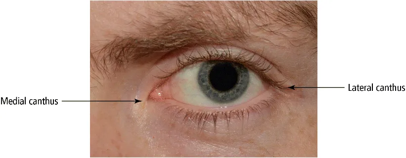
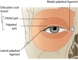
Surface Anatomy of the Face

The eyes are disposed symmetrically about the face and their forward-looking arrangement permits a large overlap in visual fields, the basis of stereopsis. Lying within the bony orbits, they are protected from trauma by the orbital walls and rims and by the eyelids, by blinking and eye closure. With the eyes open and looking straight ahead, all but the upper and lower corneal margins are exposed in the palpebral aperture, together with two small white triangles of bulbar conjunctiva, overlying the sclera. The medial and lateral ends of the fissure are known as the medial and lateral canthi (Figure 1.1).
Photograph depicting the human eye looking straight ahead. Medial canthus and lateral canthus are denoted by arrows.
Figure 1.1 The eye, looking straight ahead.
The lids and the upper and lower orbital rims are overlain by the orbicularis muscle which sweeps over these structures in an ellipse, from a region just medial to the medial orbital rim. It acts as the palpebral sphincter (Figure 1.2). Like all other muscles of the face, it is supplied by the seventh cranial nerve. Contraction of its orbital part results in protective, forced eye closure, while contraction of its palpebral part is employed in the downstroke of the upper lid during a blink. The levator palpebrae muscle, the elevator of the upper lid (see below), is concerned with the upstroke of the blink (third cranial nerve). These synchronized contractions are completed within just 300 ms. The contents of the orbit are separated from those of the lid by a connective tissue sheet, or orbital septum, which extends from the orbital rim to the tarsal plate, deep to orbicularis.
Figure depicting the diagram of human eye and its surrounding areas: medial palpebral ligament, orbicularis oculi muscle (orbital- and palpebral parts) and lateral palpebral ligament.
Figure 1.2 Disposition of the orbicularis is muscle.

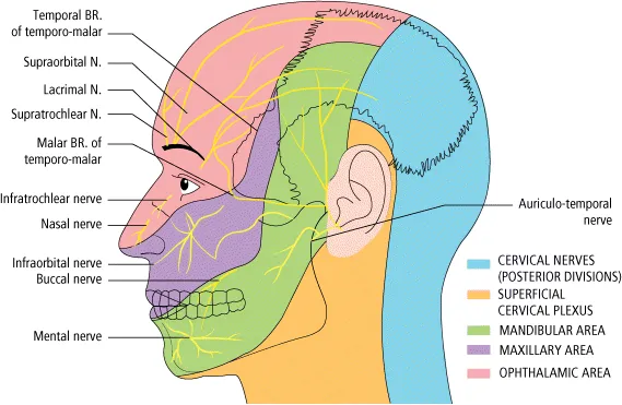
Sensory Innervation of the Face: The Fifth Cranial Nerve

The sensory innervation of each half of the face is provided by the trigeminal nerve (Figure 1.3). The eye, upper lid, eyebrow, forehead and nose are supplied by its ophthalmic division (V1), via its lacrimal, frontal and nasociliary branches, which enter the orbit through the superior orbital fissure. The maxillary division (V2), lying inferolaterally to V1 in the cavernous sinus, exits the cranial cavity via the foramen rotundum and, at the inferior orbital fissure, gives rise to the infraorbital and zygomatic nerves. These supply, chiefly, the lower lid and the upper lip and cheek. The mandibular division (V3), exiting the skull via the foramen ovale, supplies the lower lip, chin and jaw and the preauricular skin and temporal region. It is also motor to the muscles of mastication.
img
Figure 1.3 Sensory innervation of the face by the trigeminal nerve.
The neurons of the three divisions of the trigeminal nerve converge upon the trigeminal ganglion, whose sensory roots enter the pons to be distributed to the trigeminal nuclei in the brainstem. The mesencephalic nucleus is concerned with proprioception, the main sensory nucleus with touch and the medullary nucleus of the spinal tract with pain and temperature sensibility. Fibres from the ophthalmic division go to the lowest part of this nucleus, those from the mandibular division to its highest part.

Gross Anatomy of the Eye

The eye comprises (Figure 1.4):
  • A tough, collagenous outer coat which is transparent anteriorly (the cornea) and opaque posteriorly (the sclera). The junction between them is called the limbus. The extraocular muscles attach to the outer sclera, while the optic nerve leaves the globe posteriorly.
  • A rich vascular coat (the uvea) forms the choroid posteriorly and the ciliary body and iris anteriorly. Internal to the choroid lies the retina, to which it is firmly attached and whose outer two-thirds it nourishes.
  • The ciliary body contains the smooth ciliary muscle, whose contraction controls focusing by altering lens shape. The lens lies behind the iris, supported by the zonules, whose fine fibres run from the lens equator to the ciliary body. When the eye is focused for distance, tension in the zonule maintains a flattened profile of the lens. When the ciliary body contracts, tension is relaxed, the lens takes up a more ...

Inhaltsverzeichnis