An integrated, comprehensive survey of biomedical imaging modalities
An important component of the recent expansion in bioengineering is the area of biomedical imaging. This book provides in-depth coverage of the field of biomedical imaging, with particular attention to an engineering viewpoint.
Suitable as both a professional reference and as a text for a one-semester course for biomedical engineers or medical technology students, Introduction to Biomedical Imaging covers the fundamentals and applications of four primary medical imaging techniques: magnetic resonance imaging, ultrasound, nuclear medicine, and X-ray/computed tomography.
Taking an accessible approach that includes any necessary mathematics and transform methods, this book provides rigorous discussions of:
The physical principles, instrumental design, data acquisition strategies, image reconstruction techniques, and clinical applications of each modality
Recent developments such as multi-slice spiral computed tomography, harmonic and sub-harmonic ultrasonic imaging, multi-slice PET scanning, and functional magnetic resonance imaging
General image characteristics such as spatial resolution and signal-to-noise, common to all of the imaging modalities
Preguntas frecuentes
¿Cómo cancelo mi suscripción?
Simplemente, dirígete a la sección ajustes de la cuenta y haz clic en «Cancelar suscripción». Así de sencillo. Después de cancelar tu suscripción, esta permanecerá activa el tiempo restante que hayas pagado. Obtén más información aquí.
¿Cómo descargo los libros?
Por el momento, todos nuestros libros ePub adaptables a dispositivos móviles se pueden descargar a través de la aplicación. La mayor parte de nuestros PDF también se puede descargar y ya estamos trabajando para que el resto también sea descargable. Obtén más información aquí.
¿En qué se diferencian los planes de precios?
Ambos planes te permiten acceder por completo a la biblioteca y a todas las funciones de Perlego. Las únicas diferencias son el precio y el período de suscripción: con el plan anual ahorrarás en torno a un 30 % en comparación con 12 meses de un plan mensual.
¿Qué es Perlego?
Somos un servicio de suscripción de libros de texto en línea que te permite acceder a toda una biblioteca en línea por menos de lo que cuesta un libro al mes. Con más de un millón de libros sobre más de 1000 categorías, ¡tenemos todo lo que necesitas! Obtén más información aquí.
¿Perlego ofrece la función de texto a voz?
Busca el símbolo de lectura en voz alta en tu próximo libro para ver si puedes escucharlo. La herramienta de lectura en voz alta lee el texto en voz alta por ti, resaltando el texto a medida que se lee. Puedes pausarla, acelerarla y ralentizarla. Obtén más información aquí.
¿Es Introduction to Biomedical Imaging un PDF/ePUB en línea?
Sí, puedes acceder a Introduction to Biomedical Imaging de Andrew Webb en formato PDF o ePUB, así como a otros libros populares de Medicina y Radiología, radioterapia y medicina nuclear. Tenemos más de un millón de libros disponibles en nuestro catálogo para que explores.
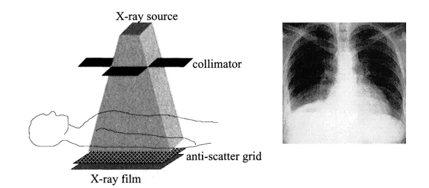
X-ray imaging is a transmission-based technique in which X-rays from a source pass through the patient and are detected either by film or an ionization chamber on the opposite side of the body, as shown in Figure 1.1. Contrast in the image between different tissues arises from differential attenuation of X-rays in the body. For example, X-ray attenuation is particularly efficient in bone, but less so in soft tissues. In planar X-ray radiography, the image produced is a simple two-dimensional projection of the tissues lying between the X-ray source and the film. Planar X-ray radiography is used for a number of different purposes: intravenous pyelography (IVP) to detect diseases of the genitourinary tract including kidney stones; abdominal radiography to study the liver, bladder, abdomen, and pelvis; chest radiography for diseases of the lung and broken ribs; and X-ray fluoroscopy (in which images are acquired continuously over a period of several minutes) for a number of different genitourinary and gastrointestinal diseases.
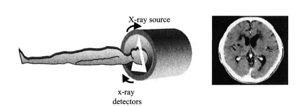
Planar X-ray radiography of overlapping layers of soft tissue or complex bone structures can often be difficult to interpret, even for a skilled radiologist. In these cases, X-ray computed tomography (CT) is used. The basic principles of CT are shown in Figure 1.2. The X-ray source is tightly collimated to interrogate a thin “slice” through the patient. The source and detectors rotate together around the patient, producing a series of one-dimensional projections at a number of different angles. These data are reconstructed to give a two-dimensional image, as shown on the right of Figure 1.2. CT images have a very high spatial resolution (~1 mm) and provide reasonable contrast between soft tissues. In addition to anatomical imaging, CT is the imaging method that can produce the highest resolution angiographic images, that is, images that show blood flow in vessels. Recent developments in spiral and multislice CT have enabled the acquisition of full three-dimensional images in a single patient breath-hold.
FIGURE 1.1. (Left) The basic setup for X-ray imaging. The collimator restricts the beam of X-rays so as to irradiate only the region of interest. The antiscatter grid increases tissue contrast by reducing the number of detected X-rays that have been scattered by tissue. (Right) A typical planar X-ray radiograph of the chest, in which the highly attenuating regions of bone appear white.
The major disadvantage of both X-ray and CT imaging is the fact that the technique uses ionizing radiation. Because ionizing radiation can cause tissue damage, there is a limit on the total radiation dose per year to which a patient can be subjected. Radiation dose is of particular concern in pediatric and obstetric radiology.
1.2. X-RAY PRODUCTION
The X-ray source is the most important system component in determining the overall image quality. Although the basic design has changed little since the mid-1900s, there have been considerable advances in the past two decades in designing more efficient X-ray sources, which are capable of delivering the much higher output levels necessary for techniques such as CT and X-ray fluoroscopy.
FIGURE 1.2. (Left) The principle of computed tomography with an X-ray source and detector unit rotating synchronously around the patient. Data are essentially acquired continuously during rotation. (Right) An example of a single-slice CT image of the brain.
1.2.1. The X-Ray Source
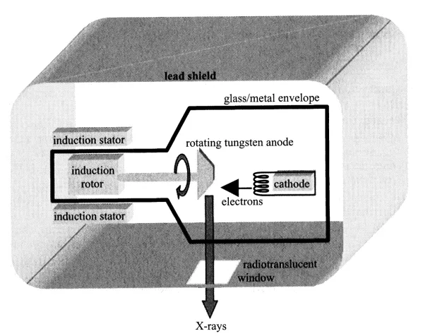
The basic components of the X-ray source, also referred to as the X-ray tube, used for clinical diagnoses are shown in Figure 1.3. The production of X-rays involves accelerating a beam of electrons to strike the surface of a metal target. The X-ray tube has two electrodes, a negatively charged cathode, which acts as the electron source, and a positively charged anode, which contains the metal target. A potential difference of between 15 and 150 kV is applied between the cathode and the anode; the exact value depends upon the particular application. This potential difference is in the form of a rectified alternating voltage, which is characterized by its maximum value, the kilovolts peak (kVp). The maximum value of the voltage is also referred to as the accelerating voltage. The cathode consists of a filament of tungsten wire (~200 μm in diameter) coiled to form a spiral ~2 mm in diameter and less than 1 cm in height. An electric current from a power source passes through the cathode, causing it to heat up. When the cathode temperature reaches ~2200°C the thermal energy absorbed by the tungsten atoms allows a small number of electrons to move away from the metallic surface, a process termed thermionic emission. A dynamic equilibrium is set up, with electrons having sufficient energy to escape from the surface of the cathode, but also being attracted back to the metal surface.
FIGURE 1.3. A schematic of an X-ray source used for clinical imaging.
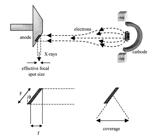
FIGURE 1.4. (Top) A negatively charged focusing cup within the X-ray cathode produces a tightly focused beam of electrons and increases the electron flux striking the tungsten anode. (Bottom) The effect of the anode bevel angleθon the effective focal spot size f and the X-ray coverage.
The large positive voltage applied to the anode causes these fre...