An integrated, comprehensive survey of biomedical imaging modalities
An important component of the recent expansion in bioengineering is the area of biomedical imaging. This book provides in-depth coverage of the field of biomedical imaging, with particular attention to an engineering viewpoint.
Suitable as both a professional reference and as a text for a one-semester course for biomedical engineers or medical technology students, Introduction to Biomedical Imaging covers the fundamentals and applications of four primary medical imaging techniques: magnetic resonance imaging, ultrasound, nuclear medicine, and X-ray/computed tomography.
Taking an accessible approach that includes any necessary mathematics and transform methods, this book provides rigorous discussions of:
The physical principles, instrumental design, data acquisition strategies, image reconstruction techniques, and clinical applications of each modality
Recent developments such as multi-slice spiral computed tomography, harmonic and sub-harmonic ultrasonic imaging, multi-slice PET scanning, and functional magnetic resonance imaging
General image characteristics such as spatial resolution and signal-to-noise, common to all of the imaging modalities
Häufig gestellte Fragen
Wie kann ich mein Abo kündigen?
Gehe einfach zum Kontobereich in den Einstellungen und klicke auf „Abo kündigen“ – ganz einfach. Nachdem du gekündigt hast, bleibt deine Mitgliedschaft für den verbleibenden Abozeitraum, den du bereits bezahlt hast, aktiv. Mehr Informationen hier.
(Wie) Kann ich Bücher herunterladen?
Derzeit stehen all unsere auf Mobilgeräte reagierenden ePub-Bücher zum Download über die App zur Verfügung. Die meisten unserer PDFs stehen ebenfalls zum Download bereit; wir arbeiten daran, auch die übrigen PDFs zum Download anzubieten, bei denen dies aktuell noch nicht möglich ist. Weitere Informationen hier.
Welcher Unterschied besteht bei den Preisen zwischen den Aboplänen?
Mit beiden Aboplänen erhältst du vollen Zugang zur Bibliothek und allen Funktionen von Perlego. Die einzigen Unterschiede bestehen im Preis und dem Abozeitraum: Mit dem Jahresabo sparst du auf 12 Monate gerechnet im Vergleich zum Monatsabo rund 30 %.
Was ist Perlego?
Wir sind ein Online-Abodienst für Lehrbücher, bei dem du für weniger als den Preis eines einzelnen Buches pro Monat Zugang zu einer ganzen Online-Bibliothek erhältst. Mit über 1 Million Büchern zu über 1.000 verschiedenen Themen haben wir bestimmt alles, was du brauchst! Weitere Informationen hier.
Unterstützt Perlego Text-zu-Sprache?
Achte auf das Symbol zum Vorlesen in deinem nächsten Buch, um zu sehen, ob du es dir auch anhören kannst. Bei diesem Tool wird dir Text laut vorgelesen, wobei der Text beim Vorlesen auch grafisch hervorgehoben wird. Du kannst das Vorlesen jederzeit anhalten, beschleunigen und verlangsamen. Weitere Informationen hier.
Ist Introduction to Biomedical Imaging als Online-PDF/ePub verfügbar?
Ja, du hast Zugang zu Introduction to Biomedical Imaging von Andrew Webb im PDF- und/oder ePub-Format sowie zu anderen beliebten Büchern aus Medicina & Radiología, radioterapia y medicina nuclear. Aus unserem Katalog stehen dir über 1 Million Bücher zur Verfügung.
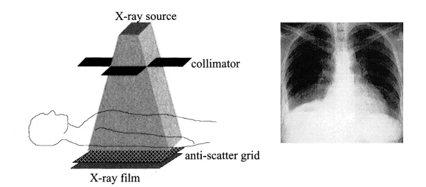
X-ray imaging is a transmission-based technique in which X-rays from a source pass through the patient and are detected either by film or an ionization chamber on the opposite side of the body, as shown in Figure 1.1. Contrast in the image between different tissues arises from differential attenuation of X-rays in the body. For example, X-ray attenuation is particularly efficient in bone, but less so in soft tissues. In planar X-ray radiography, the image produced is a simple two-dimensional projection of the tissues lying between the X-ray source and the film. Planar X-ray radiography is used for a number of different purposes: intravenous pyelography (IVP) to detect diseases of the genitourinary tract including kidney stones; abdominal radiography to study the liver, bladder, abdomen, and pelvis; chest radiography for diseases of the lung and broken ribs; and X-ray fluoroscopy (in which images are acquired continuously over a period of several minutes) for a number of different genitourinary and gastrointestinal diseases.
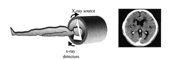
Planar X-ray radiography of overlapping layers of soft tissue or complex bone structures can often be difficult to interpret, even for a skilled radiologist. In these cases, X-ray computed tomography (CT) is used. The basic principles of CT are shown in Figure 1.2. The X-ray source is tightly collimated to interrogate a thin “slice” through the patient. The source and detectors rotate together around the patient, producing a series of one-dimensional projections at a number of different angles. These data are reconstructed to give a two-dimensional image, as shown on the right of Figure 1.2. CT images have a very high spatial resolution (~1 mm) and provide reasonable contrast between soft tissues. In addition to anatomical imaging, CT is the imaging method that can produce the highest resolution angiographic images, that is, images that show blood flow in vessels. Recent developments in spiral and multislice CT have enabled the acquisition of full three-dimensional images in a single patient breath-hold.
FIGURE 1.1. (Left) The basic setup for X-ray imaging. The collimator restricts the beam of X-rays so as to irradiate only the region of interest. The antiscatter grid increases tissue contrast by reducing the number of detected X-rays that have been scattered by tissue. (Right) A typical planar X-ray radiograph of the chest, in which the highly attenuating regions of bone appear white.
The major disadvantage of both X-ray and CT imaging is the fact that the technique uses ionizing radiation. Because ionizing radiation can cause tissue damage, there is a limit on the total radiation dose per year to which a patient can be subjected. Radiation dose is of particular concern in pediatric and obstetric radiology.
1.2. X-RAY PRODUCTION
The X-ray source is the most important system component in determining the overall image quality. Although the basic design has changed little since the mid-1900s, there have been considerable advances in the past two decades in designing more efficient X-ray sources, which are capable of delivering the much higher output levels necessary for techniques such as CT and X-ray fluoroscopy.
FIGURE 1.2. (Left) The principle of computed tomography with an X-ray source and detector unit rotating synchronously around the patient. Data are essentially acquired continuously during rotation. (Right) An example of a single-slice CT image of the brain.
1.2.1. The X-Ray Source
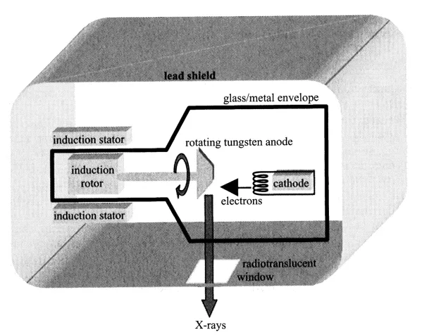
The basic components of the X-ray source, also referred to as the X-ray tube, used for clinical diagnoses are shown in Figure 1.3. The production of X-rays involves accelerating a beam of electrons to strike the surface of a metal target. The X-ray tube has two electrodes, a negatively charged cathode, which acts as the electron source, and a positively charged anode, which contains the metal target. A potential difference of between 15 and 150 kV is applied between the cathode and the anode; the exact value depends upon the particular application. This potential difference is in the form of a rectified alternating voltage, which is characterized by its maximum value, the kilovolts peak (kVp). The maximum value of the voltage is also referred to as the accelerating voltage. The cathode consists of a filament of tungsten wire (~200 μm in diameter) coiled to form a spiral ~2 mm in diameter and less than 1 cm in height. An electric current from a power source passes through the cathode, causing it to heat up. When the cathode temperature reaches ~2200°C the thermal energy absorbed by the tungsten atoms allows a small number of electrons to move away from the metallic surface, a process termed thermionic emission. A dynamic equilibrium is set up, with electrons having sufficient energy to escape from the surface of the cathode, but also being attracted back to the metal surface.
FIGURE 1.3. A schematic of an X-ray source used for clinical imaging.
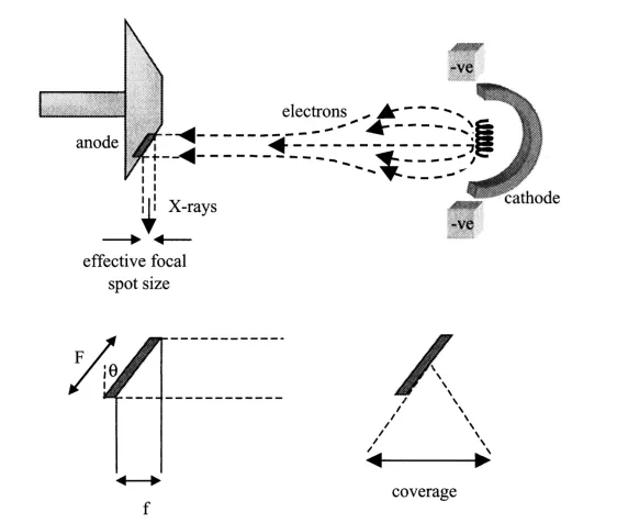
FIGURE 1.4. (Top) A negatively charged focusing cup within the X-ray cathode produces a tightly focused beam of electrons and increases the electron flux striking the tungsten anode. (Bottom) The effect of the anode bevel angleθon the effective focal spot size f and the X-ray coverage.
The large positive voltage applied to the anode causes these fre...