Veterinary Microbiology and Microbial Disease
P. J. Quinn, B. K. Markey, F. C. Leonard, P. Hartigan, S. Fanning, E. S. Fitzpatrick
- English
- ePUB (apto para móviles)
- Disponible en iOS y Android
Veterinary Microbiology and Microbial Disease
P. J. Quinn, B. K. Markey, F. C. Leonard, P. Hartigan, S. Fanning, E. S. Fitzpatrick
Información del libro
Microbiology is one of the core subjects for veterinary students, and since its first publication in 2002, Veterinary Microbiology and Microbial Disease has become an essential text for students of veterinary medicine. Fully revised and expanded, this new edition updates the subject for pre-clinical and clinical veterinary students in a comprehensive manner. Individual sections deal with bacteriology, mycology and virology. Written by an academic team with many years of teaching experience, the book provides concise descriptions of groups of microorganisms and the diseases which they cause. Microbial pathogens are discussed in separate chapters which provide information on the more important features of each microorganism and its role in the pathogenesis of diseases of animals. The international and public health significance of these pathogens are reviewed comprehensively. The final section is concerned with the host and is organized according to the body system affected.
Tables, boxes and flow diagrams provide information in an easily assimilated format. This edition contains new chapters on molecular diagnostics and on infectious conditions of the skin, cardiovascular system, urinary tract and musculoskeletal system. Many new colour diagrams are incorporated into this edition and each chapter has been updated.
Key features of this edition:
- Twelve new chapters included
- Numerous new illustrations
- Each chapter has been updated
- Completely re-designed in full colour
- Fulfils the needs of veterinary students and academics in veterinary microbiology
- Companion website with figures from the book as Powerpoints for viewing or downloading by chapter: www.wiley.com/go/quinn/veterinarymicrobiology
Veterinary Microbiology and Microbial Disease remains indispensable for all those studying and teaching this essential component of the veterinary curriculum.
Preguntas frecuentes
Información
Pathogenic Bacteria
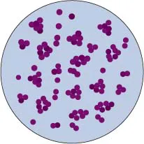
- Gram-positive cocci in clusters resembling bunches of grapes
- Grow on non-enriched media
- Moderately-sized white or golden colonies
- Colonies of S. aureus and S. pseudintermedius produce double haemolysis
- Facultative anaerobes, non-motile, catalase-positive
- Commensals on mucous membranes and skin
- Coagulase production correlates with pathogenicity
- Comparatively stable in the environment
- Cause pyogenic infections
| Species | Hosts | Clinical conditions |
| Staphylococcus aureusa | Cattle | Mastitis, udder impetigo |
| Sheep | Mastitis | |
| Tick pyaemia (lambs) | ||
| Benign folliculitis (lambs) | ||
| Dermatitis | ||
| Goats | Mastitis | |
| Dermatitis | ||
| Pigs | Botryomycosis of mammary glands | |
| Impetigo on mammary glands | ||
| Horses | Scirrhous cord (botryomycosis of the spermatic cord), mastitis | |
| Dogs, cats | Suppurative conditions similar to those caused by S. pseudintermedius | |
| Poultry | Arthritis and septicaemia in turkeys | |
| Bumblefoot | ||
| Omphalitis in chicks | ||
| S. pseudintermedius | Dogs | Pyoderma, endometritis, cystitis, otitis externa, and other suppurative conditions |
| Cats | Various pyogenic conditions | |
| Horses | Rarely isolated | |
| Cows | Rarely isolated | |
| S. hyicusb | Pigs | Exudative epidermitis (greasy-pig disease) |
| Arthritis | ||
| Cattle | Mastitis (rare) | |
| S. intermedius | Horses | Isolated from nares |
| Pigeons | Isolated from upper respiratory tract | |
| S. aureus subsp. anaerobius | Sheep | Lymphadenitis |
| S. delphini | Dolphins | Suppurative skin lesions |
| Horses | Isolated from nares | |
| Pigeons | Isolated from upper respiratory tract | |
| S. lutrae | Otters | Pathogenic significance uncertain |
| S. schleiferi subsp. coagulans | Dogs | Otitis externa |
| Species | Host/Source |
| S. arlettae | Goats/Nares |
| Poultry/Skin | |
| S. capitis | Cattle/Milk |
| S. caprae | Goats/Skin |
| S. chromogenes | Cattle/Milka |
| Pigs, poultry/Skin | |
| S. cohnii | Cattle/Milka |
| S. epidermidis | Cattle/Milka |
| Dogs, horses/Wound infections | |
| S. equorum | Horses/Skin |
| S. felisb | Cats/Otitis externa, skin infections |
| S. gallinarum | Poultry/Skin infections |
| S. haemolyticus | Cattle/Milka |
| S. hominis | Cattle/Milk |
| S. lentus | Pigs, sheep, goats/Skin infections |
| S. nepalensis | Goats/Respiratory tract |
| S. saprophyticus | Cats/Skin |
| Cattle/Nostrils | |
| S. sciuri | Cats and other animals/Skin infections |
| S. simiae | Squirrel monkeys/Gastrointestinal tract |
| S. simulans | Cattle/Milka |
| Dogs, cats, pigs/Skin | |
| S. vitulinus | Cattle, sheep, pigs/Skin |
| S. warneri | Cattle/Milka |
| S. xylosus | Cattle, sheep/Milka |
| Cats, poultry, pigs, horses/Skin |
- Colonial characteristics: Staphylococcal colonies are usually white, opaque and up to 4 mm in diameter. The colonies of bovine and human strains of S. aureus are golden yellow. Colonies of some coagulase-negative staphylococci are also pigmented.
- Haemolysis in sheep or ox blood agar: Four staphylococcal haemolysins are recognized, alpha, beta, gamma and delta. Individual haemolysins differ antigenically, biochemically and in their effects on the red blood cells of different animal species. Strains vary in their haemolysin-producing ability, and animal strains of S. aureus and S. pseudintermedius usually produce both alpha-haemolysin and beta-haemolysin. On ruminant blood agar, the alpha-haemolysin causes a narrow zone of complete haemolysis immediately around the colony, and the beta-haemolysin produces a wider zone of partial or incomplete haemolysis. This is referred to as double haemolysis (Fig. 14.2). These haemolysins act as toxins in vivo. Coagulase-negative staphylococci exhibit variation in their ability to produce haemolysis which usually develops slowly. Isolates of S. hyicus are non-haemolytic.
- Slide and tube coagulase tests: In these tests, a suspension of staphylococci is mixed with rabbit plasma either on a slide or in a small tube. The fibrinogen in rabbit plasma i...