Chapter 1
The Helicobacter Genus: The History of H. Pylori and Taxonomy of Current Species
C. Stewart Goodwin and Bryan W. Worsley
TABLE OF CONTENTS
I. Introduction
II. History of H. pylori
A. Early Papers
B. First Culture of H. pylori in 1982 and Realization of the New Genus
III. Taxonomic Features of the Helicobacter Genus
A. Ultrastructure
B. Growth Conditions
C. Biochemical Capabilities
D. Antibiotic Susceptibility
E. Cellular Fatty Acid Composition
F. Menaquinones and Lipid A
G. Phylogenetic Analysis
1. DNA Base Composition
2. Hybridization
3. RNA Sequencing
H. Protein Analysis
I. Immunoanalysis
IV. Conclusion
References
I. Introduction
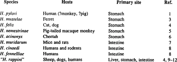
How the name Helicobacter was chosen is described in the Preface; the genus name was first published in 1989, with two species, H. pylori and H. mustelae.1 Teamwork, among four departments, was the secret of the first successful culture of H. pylori in Royal Perth Hospital, Western Australia, in 19822 (see below). By 1992, this new genus contained nine named species (Table 1) with at least four other unnamed species identified. The table shows that five species are gastric pathogens, three other species are lower gut organisms, and “H. rappini” has been found in a variety of sites. “H. rappini” was originally named “Flexispira rappini”;9 although it has some phenotypic differences from H. pylori,’ it possesses urease and genotypically should be in the Helicobacter genus.4, 8 “H. rappini” has been isolated from the widest range of hosts and tissues. It was first isolated from the liver of aborted lambs,10then seen in the stomachs of beagle dogs,11 and then isolated from feces of humans with gastroenteritis.12 Helicobacter species have been found in the feces of terns, gulls, house sparrows, and pigs, and a Helicobacter species distinct from H. mustelae has been found in ferret feces (Paster, personal communication). “Gastrospirillum hominis”,13 which has not been cultured nor characterized, has not been officially named. However, RNA sequencing has shown that “G. hominis” is also in the Helicobacter genus, and the name “H. heilmanii” has been suggested.13®
II. History Of H. Pylori
A. Early Papers
Credit for the first description of spiral organisms in the human stomach should probably be given to Bottcher in 1974.14 Bizzozero in 189315 and Salomon in 189616 described “spirochaetes” in animals, but these were unlikely to have been H. pylori. Probably they were “H. heilmanii.” Krienitz described human gastric spiral organisms in 1906,17 and Freedberg and Barron in 1940.18 Those who described spiral bacteria in humans and animals usually failed to distinguish the shorter, gently spiraled H. pylori from the longer, tightly spiraled “H. heilmanii.”19 Even in 1987, when “H. rappini” was noted in beagles, the three electron micrographs in fact showed ”H. rappini,” H. felis, and “H. heilmanii.”11 H. muridarum was cultured in Sydney about the same time as H. pylori.20 However, it was officially named only in January 1992.7 Studies of human H. pylori were at first only histological and ultrastructural, without successful culture. In 1975, Steer noted that spiral bacteria were closely apposed to the gastric mucus secreting cells,21 but culture yielded only Pseudomonas aeruginosa.22 Electron micrographs of spiral bacteria in large numbers on gastric epithelial cells were published by him in May 1984.23Independently, in Birmingham, England, in 1981 Rollason et al. had observed gastric spiral bacteria.24
TABLE 1
Helicobacter Species and their Hosts (as of 1992)
B. First Culture of H. Pylori in 1982 and Realization Of the new Genus
In Perth, Western Australia, at the Royal Perth Hospital, histological and ultrastructural studies of the gastric mucosa had been published in 1979;25spiral bacteria were seen, but because they did not invade the mucosa were thought to be irrelevant. The histopathologist Warren correlated them with the presence of polymorphonuclear leukocytes.26 In 1981, Marshall was training in internal medicine, and for 6 months was learning gastroenterology. With Warren he reviewed the patients in whom large numbers of gastric spiral bacteria had been seen.27 One of these had been treated fortuitously with tetracycline; his symptoms resolved, and subsequent endoscopic biopsy showed that the antral gastritis had also resolved.27 By then Armstrong had come to Royal Perth Hospital from Mill Hill to head the Electronmicroscopy Unit at Royal Perth Hospital. He obtained high-magnification electron micrographs of H. pylori. (When Warren and Marshall could not agree on the wording of a joint letter to The Lancet in 1983, Armstrong advised them to write separate letters.26, 28) Goodwin was Head of the Microbiology Department, and in late 1981 Marshall asked him for microbiological assistance. A protocol was agreed; gastric biopsy specimens from 100 consecutive patients would be taken by the consultant gastroenterologists Waters and Sanderson and these would be processed by Gram stain and culture and by the histopathology department. The project started in March 1982, with the microbiologist Pearman supervising the project. Among the first 34 specimens, spiral bacteria were seen in the Gram stain in 6. However, in spite of frequent variations of media, and incubation temperatures, spiral bacteria were not cultured, because incubation was limited to 48 h. The 35th culture was left incubating during the Easter holiday, which in Australia lasted for 5 d. When the plates were finally viewed, a pure growth of 1-mm transparent colonies were seen. H. pylori had been finally cultured! The date was the 14th of April, 1982.2 Fro...