Marie Svoboda
Associate Conservator, Antiquities Conservation, J. Paul Getty Museum, Los Angeles, CA
C. Michael Bowers
Associate Clinical Professor, Herman Ostrow School of Dentistry, University of Southern California, Los Angeles, CA
Overview
A death investigation of unidentified human remains requires professional determination of all physical evidence available from the body. The unknown person’s gender, age, medical status and cause of death are vital information for a forensic autopsy report. The following case explains details of the life and death of a young man whose body was originally found in Egypt. Dental information was important to confirm his age and give insight to his health history at the time of his death.
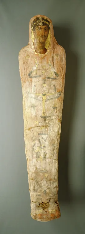
In 2003, the J. Paul Getty Museum Antiquities Conservation Department initiated the study of a Romano-Egyptian red-shroud mummy (91.AP.6) in the museum’s collection. The mummy is known as Herakleides from a painted inscription on top of the wrapped feet. Dating to the first century AD, the mummy incorporates a portrait panel depicting a young man in his early twenties. The mummy, measuring 175 cm in length, was wrapped in one large outer shroud that had been painted red and decorated with Egyptian funerary images. The beautifully executed portrait, the quality of the wrappings, and the elaborate use of gold, on both the panel and the shroud, attest to the prominent status of this individual.
The investigation of Herakleides’ mummy began with the conservation treatment of the fragile foot area, which was damaged and unstable [2]. The conservation treatment was in preparation for the mummy’s first public display at the Getty Villa in 2005, where its presence in the gallery contextualizes the now detached Romano-Egyptian portraits in the collection by illustrating their mortuary function. From here a full study of the body (human remains) and the materials used for the mummification and decoration of Herakleides evolved. The aim of this study was to better understand the person within the wrappings and the ancient techniques employed in its fabrication and adornment process. Imaging technology such as computerized tomography (CT) and infrared photographic techniques revealed secrets such as his complete name and the curious inclusion of a mummified ibis within the wrappings. Examination of the skeleton by orthopedic surgeons and a forensic dentist established his age, health, and height at the time of death. Radiocarbon dating (carbon 14) provided a secure date for the materials used in the mummification process and a likely time frame for when he lived. Motifs and religious iconography were studied and documented to better understand their meaning. The lack of clothing on the youth’s shoulders suggests he was an ephebe, or adolescent male of social standing. His presumed nudity, a symbol of rebirth, indicates he may have been an initiate in the cult of the Egyptian goddess Isis.
This study also involved the Getty Conservation Institute (GCI), which scientifically identified and compared the red pigment used on seven of the nine mummies identified within the red-shroud subgroup. The results from the analyses revealed that the composition of the unique red pigment is identical, relating this group to one another even further. The study of Herakleides shows how the collaboration of experts within the medical, scientific, and Egyptological communities can come together to better understand one unique artifact. This supportive exchange of experience, knowledge, and information has opened a window into the life, religion, and ritual of a man who lived almost 2,000 years ago [3].
The Forensic Examination of Herakleides
CT scans of the Herakleides’ mummy revealed that, contrary to the usual Egyptian practices of mummification, the 20-year-old man’s heart, not his lungs, were removed during embalming. Also uncommon in the scientists’ findings was a mummified ibis, inexplicably placed on Herakleides’ abdomen under the final layer of his mummy’s wrappings.
The Aging of Herakleides
Skeletal Analysis
His age determination was made by examining the epiphyses of his arms and legs. These are “growth plates” seen during teenage and early adult years that gradually disappear at maturity. They were faint in the Herakleides’ CT scan but were not completely fused. This is the data that produced an age range of 20 +/– 2 years. This opinion was provided by a radiologist at UCLA, who performed the CT scans, and was corroborated by two orthopaedic surgeons who examined Herakleides’ skeleton. There was no evidence of medical pathology (disease) or before-death (antemortem) trauma. A large gash is visible at the back of the skull, but it not clear whether this occurred before (antemortem) or after death (postmortem). The medical team who examined the CT scans believe it was most likely caused during mummification.
Dental Aging
Over the years, development of third molars (wisdom teeth) in adults has been researched in multiple population studies [4]. These studies compare the third molar root growth stages and development in the jawbone to the chronological (real age) of the known people in the study sample. Herekleides’ teeth are completely developed (the roots are fully formed) and are at the completed Stage H of full growth [5]. This indicates that he was at least 18 or older when he died. This supports the previous orthopaedic/anthropological opinions of this skeletal age at death.
Acknowledgments
Photo: The J. Paul Getty Museum, Villa Collection, Malibu, California
References
[1] Corcoran L. Portrait Mummies from Roman Egypt (I-IV Centuries A.D.), with a catalogue of portrait mummies in Egyptian Museums. Chicago Press, 1995;7.
[2] . http://getty.edu/art/videos/mummification_process/mummification_process.html
[3] Corcoran L., Svoboda M. Herakleides: A Red-Shroud Portrait Mummy from Roman Egypt. Los Angeles: J. Paul Getty Museum, 2010.
[4] Bowers C.M. Determining Age from Teeth: The Estimation of Age From Dental Development. In: Bowers C.M., Bell G.L., editors. Manual of Forensic Odontology. third ed. ASFO; 1995:74-105.
[5] Ibid. p. 88.