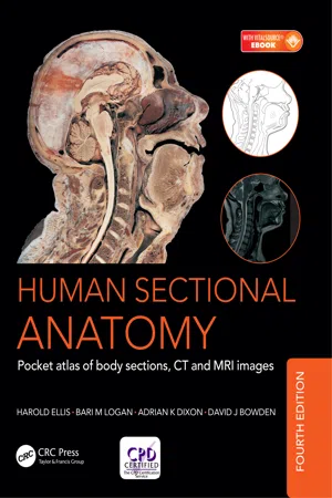
Human Sectional Anatomy
eBook - ePub

Human Sectional Anatomy

Pocket atlas of body sections, CT and MRI images, Fourth edition

Adrian Kendal Dixon, David J. Bowden, Bari M. Logan, Harold Ellis

Compartir libro
  1. 270 páginas
  2. English
  3. ePUB (apto para móviles)
  4. Disponible en iOS y Android
eBook - ePub

Human Sectional Anatomy

Pocket atlas of body sections, CT and MRI images, Fourth edition

Adrian Kendal Dixon, David J. Bowden, Bari M. Logan, Harold Ellis

Detalles del libro
Vista previa del libro
Índice
Citas

Información del libro

First published in 1991, Human Sectional Anatomy set new standards for the quality of cadaver sections and accompanying radiological images. Now in its fourth edition, this unsurpassed quality remains and is further enhanced by the addition of new material.

The superb full-colour cadaver sections are compared with CT and MRI images, with accompanying, labelled, line diagrams. Many of the radiological images have been replaced with new examples for this latest edition, captured using the most up-to date imaging technologies to ensure excellent visualization of the anatomy. The photographic material is enhanced by useful notes with details of important anatomical and radiological features.

Beautifully presented in a convenient and portable format, the fourth edition of this popular pocket atlas continues to be an essential textbook for medical and allied health students and those taking postgraduate qualifications in radiology, surgery and medicine, and an invaluable ready-reference for all practising anatomists, radiologists, radiographers, surgeons and medics.

Preguntas frecuentes

¿Cómo cancelo mi suscripción?
Simplemente, dirígete a la sección ajustes de la cuenta y haz clic en «Cancelar suscripción». Así de sencillo. Después de cancelar tu suscripción, esta permanecerá activa el tiempo restante que hayas pagado. Obtén más información aquí.
¿Cómo descargo los libros?
Por el momento, todos nuestros libros ePub adaptables a dispositivos móviles se pueden descargar a través de la aplicación. La mayor parte de nuestros PDF también se puede descargar y ya estamos trabajando para que el resto también sea descargable. Obtén más información aquí.
¿En qué se diferencian los planes de precios?
Ambos planes te permiten acceder por completo a la biblioteca y a todas las funciones de Perlego. Las únicas diferencias son el precio y el período de suscripción: con el plan anual ahorrarás en torno a un 30 % en comparación con 12 meses de un plan mensual.
¿Qué es Perlego?
Somos un servicio de suscripción de libros de texto en línea que te permite acceder a toda una biblioteca en línea por menos de lo que cuesta un libro al mes. Con más de un millón de libros sobre más de 1000 categorías, ¡tenemos todo lo que necesitas! Obtén más información aquí.
¿Perlego ofrece la función de texto a voz?
Busca el símbolo de lectura en voz alta en tu próximo libro para ver si puedes escucharlo. La herramienta de lectura en voz alta lee el texto en voz alta por ti, resaltando el texto a medida que se lee. Puedes pausarla, acelerarla y ralentizarla. Obtén más información aquí.
¿Es Human Sectional Anatomy un PDF/ePUB en línea?
Sí, puedes acceder a Human Sectional Anatomy de Adrian Kendal Dixon, David J. Bowden, Bari M. Logan, Harold Ellis en formato PDF o ePUB, así como a otros libros populares de Medicine y Medical Theory, Practice & Reference. Tenemos más de un millón de libros disponibles en nuestro catálogo para que explores.

Información

Editorial
CRC Press
Año
2017
ISBN
9781498708708

Contents

Preface
Introduction
The importance of cross-sectional anatomy
Orientation of sections and images
Notes on the atlas
References
Acknowledgments
Interpreting cross-sections: helpful hints for medical students
➜ BRAIN
Series of Superficial Dissections [A–H]
➜ HEAD
Base of skull [Osteology]
Cranial fossae [Cranial nerves dissection]
Sagittal section
Sagittal section [Cranial nerves dissection]
Axial sections [1–19 Male]
Selected images
Axial Magnetic Resonance Images [A–C]
Coronal sections [1–13 Female]
Sagittal section [1 Male]
TEMPORAL BONE/INNER EAR
Coronal sections [1–2 Male]
Selected images
Axial Computed Tomogram [A] Temporal Bone/Inner Ear
➜ NECK
Axial sections [1–9 Female]
Sagittal section [1 Male]
➜ THORAX
Axial sections [1–10 Male]
Axial section [1 Female]
Selected images
Axial Computed Tomograms [A–C] Heart
Axial Computed Tomograms [A–D] Mediastinum
Coronal Magnetic Resonance Images [A–C]
Reconstructed Computed Tomograms [A–E] Chest
Reconstructed 3D Computed Tomograms [A–B] Arterial System
➜ ABDOMEN
Axial sections [1–8 Male]
Axial sections [1–2 Female]
Selected images
3D Computed Tomography Colonogram [A]
Axial Computed Tomograms [A–F] Lumbar Spine
Coronal Magnetic Resonance Images [A–B] Lumbar Spine
Sagittal Magnetic Resonance Images [A–D] Lumbar Spine
➜ PELVIS
Axial sections [1–11 Male]
Selected images
Coronal Magnetic Resonance Images [A–C]
Axial sections [1–7 Female]
Selected images
Axial Magnetic Resonance Images [A–B]
Coronal Magnetic Resonance Images [A–C]
Sagittal Magnetic Resonance Image [A]
➜ LOWER LIMB
HIP – Coronal section [1 Female]
Selected images
3D Computed Tomograms [A–B] Pelvis
THIGH – Axial sections [1–3 Male]
KNEE – Axial sections [1–3 Male]
KNEE – Coronal section [1 Male]
KNEE – Sagittal sections [1–3 Female]
LEG – Axial sections [1–2 Male]
ANKLE – Axial sections [1–3 Male]
ANKLE – Coronal section [1 Female]
ANKLE/FOOT – Sagittal section [1 Male]
FOOT – Coronal section [1 Male]
➜ UPPER LIMB
SHOULDER – Axial section [1 Female]
SHOULDER – Coronal section [1 Male]
Selected images
3D Computed Tomograms [A–B] Shoulder Girdle
ARM – Axial section [1 Male]
ELBOW – Axial sections [1–3 Male]
ELBOW – Coronal section [1 Female]
FOREARM – Axial sections [1–2 Male]
WRIST – Axial sections [1–3 Male]
WRIST/HAND – Coronal section [1 Female]
WRIST/HAND – Sagittal section [1 Female]
HAND – Axial sections [1–2 Male]
Index

Preface

The study of sectional anatomy of the human body goes back to the earliest days of systematic topographical anatomy. The beautiful drawings of the sagittal sections of the male and female trunk and of the pregnant uterus by Leonardo da Vinci (1452-1519) are well known. Among his figures, which were based on some 30 dissections, are a number of transverse sections of the lower limb. These constitute the first known examples of the use of cross-sections for the study of gross anatomy and anticipate modern technique by several hundred years. In the absence of...

Índice