Human Sectional Anatomy
eBook - ePub

Human Sectional Anatomy

Pocket atlas of body sections, CT and MRI images, Fourth edition

Adrian Kendal Dixon, David J. Bowden, Bari M. Logan, Harold Ellis

Buch teilen
  1. 270 Seiten
  2. English
  3. ePUB (handyfreundlich)
  4. Über iOS und Android verfĂŒgbar
eBook - ePub

Human Sectional Anatomy

Pocket atlas of body sections, CT and MRI images, Fourth edition

Adrian Kendal Dixon, David J. Bowden, Bari M. Logan, Harold Ellis

Angaben zum Buch
Buchvorschau
Inhaltsverzeichnis
Quellenangaben

Über dieses Buch

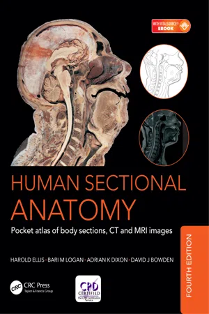
First published in 1991, Human Sectional Anatomy set new standards for the quality of cadaver sections and accompanying radiological images. Now in its fourth edition, this unsurpassed quality remains and is further enhanced by the addition of new material.

The superb full-colour cadaver sections are compared with CT and MRI images, with accompanying, labelled, line diagrams. Many of the radiological images have been replaced with new examples for this latest edition, captured using the most up-to date imaging technologies to ensure excellent visualization of the anatomy. The photographic material is enhanced by useful notes with details of important anatomical and radiological features.

Beautifully presented in a convenient and portable format, the fourth edition of this popular pocket atlas continues to be an essential textbook for medical and allied health students and those taking postgraduate qualifications in radiology, surgery and medicine, and an invaluable ready-reference for all practising anatomists, radiologists, radiographers, surgeons and medics.

HĂ€ufig gestellte Fragen

Wie kann ich mein Abo kĂŒndigen?
Gehe einfach zum Kontobereich in den Einstellungen und klicke auf „Abo kĂŒndigen“ – ganz einfach. Nachdem du gekĂŒndigt hast, bleibt deine Mitgliedschaft fĂŒr den verbleibenden Abozeitraum, den du bereits bezahlt hast, aktiv. Mehr Informationen hier.
(Wie) Kann ich BĂŒcher herunterladen?
Derzeit stehen all unsere auf MobilgerĂ€te reagierenden ePub-BĂŒcher zum Download ĂŒber die App zur VerfĂŒgung. Die meisten unserer PDFs stehen ebenfalls zum Download bereit; wir arbeiten daran, auch die ĂŒbrigen PDFs zum Download anzubieten, bei denen dies aktuell noch nicht möglich ist. Weitere Informationen hier.
Welcher Unterschied besteht bei den Preisen zwischen den AboplÀnen?
Mit beiden AboplÀnen erhÀltst du vollen Zugang zur Bibliothek und allen Funktionen von Perlego. Die einzigen Unterschiede bestehen im Preis und dem Abozeitraum: Mit dem Jahresabo sparst du auf 12 Monate gerechnet im Vergleich zum Monatsabo rund 30 %.
Was ist Perlego?
Wir sind ein Online-Abodienst fĂŒr LehrbĂŒcher, bei dem du fĂŒr weniger als den Preis eines einzelnen Buches pro Monat Zugang zu einer ganzen Online-Bibliothek erhĂ€ltst. Mit ĂŒber 1 Million BĂŒchern zu ĂŒber 1.000 verschiedenen Themen haben wir bestimmt alles, was du brauchst! Weitere Informationen hier.
UnterstĂŒtzt Perlego Text-zu-Sprache?
Achte auf das Symbol zum Vorlesen in deinem nÀchsten Buch, um zu sehen, ob du es dir auch anhören kannst. Bei diesem Tool wird dir Text laut vorgelesen, wobei der Text beim Vorlesen auch grafisch hervorgehoben wird. Du kannst das Vorlesen jederzeit anhalten, beschleunigen und verlangsamen. Weitere Informationen hier.
Ist Human Sectional Anatomy als Online-PDF/ePub verfĂŒgbar?
Ja, du hast Zugang zu Human Sectional Anatomy von Adrian Kendal Dixon, David J. Bowden, Bari M. Logan, Harold Ellis im PDF- und/oder ePub-Format sowie zu anderen beliebten BĂŒchern aus Medicine & Medical Theory, Practice & Reference. Aus unserem Katalog stehen dir ĂŒber 1 Million BĂŒcher zur VerfĂŒgung.

Information

Verlag
CRC Press
Jahr
2017
ISBN
9781498708708

Contents

Preface
Introduction
The importance of cross-sectional anatomy
Orientation of sections and images
Notes on the atlas
References
Acknowledgments
Interpreting cross-sections: helpful hints for medical students
➜ BRAIN
Series of Superficial Dissections [A–H]
➜ HEAD
Base of skull [Osteology]
Cranial fossae [Cranial nerves dissection]
Sagittal section
Sagittal section [Cranial nerves dissection]
Axial sections [1–19 Male]
Selected images
Axial Magnetic Resonance Images [A–C]
Coronal sections [1–13 Female]
Sagittal section [1 Male]
TEMPORAL BONE/INNER EAR
Coronal sections [1–2 Male]
Selected images
Axial Computed Tomogram [A] Temporal Bone/Inner Ear
➜ NECK
Axial sections [1–9 Female]
Sagittal section [1 Male]
➜ THORAX
Axial sections [1–10 Male]
Axial section [1 Female]
Selected images
Axial Computed Tomograms [A–C] Heart
Axial Computed Tomograms [A–D] Mediastinum
Coronal Magnetic Resonance Images [A–C]
Reconstructed Computed Tomograms [A–E] Chest
Reconstructed 3D Computed Tomograms [A–B] Arterial System
➜ ABDOMEN
Axial sections [1–8 Male]
Axial sections [1–2 Female]
Selected images
3D Computed Tomography Colonogram [A]
Axial Computed Tomograms [A–F] Lumbar Spine
Coronal Magnetic Resonance Images [A–B] Lumbar Spine
Sagittal Magnetic Resonance Images [A–D] Lumbar Spine
➜ PELVIS
Axial sections [1–11 Male]
Selected images
Coronal Magnetic Resonance Images [A–C]
Axial sections [1–7 Female]
Selected images
Axial Magnetic Resonance Images [A–B]
Coronal Magnetic Resonance Images [A–C]
Sagittal Magnetic Resonance Image [A]
➜ LOWER LIMB
HIP – Coronal section [1 Female]
Selected images
3D Computed Tomograms [A–B] Pelvis
THIGH – Axial sections [1–3 Male]
KNEE – Axial sections [1–3 Male]
KNEE – Coronal section [1 Male]
KNEE – Sagittal sections [1–3 Female]
LEG – Axial sections [1–2 Male]
ANKLE – Axial sections [1–3 Male]
ANKLE – Coronal section [1 Female]
ANKLE/FOOT – Sagittal section [1 Male]
FOOT – Coronal section [1 Male]
➜ UPPER LIMB
SHOULDER – Axial section [1 Female]
SHOULDER – Coronal section [1 Male]
Selected images
3D Computed Tomograms [A–B] Shoulder Girdle
ARM – Axial section [1 Male]
ELBOW – Axial sections [1–3 Male]
ELBOW – Coronal section [1 Female]
FOREARM – Axial sections [1–2 Male]
WRIST – Axial sections [1–3 Male]
WRIST/HAND – Coronal section [1 Female]
WRIST/HAND – Sagittal section [1 Female]
HAND – Axial sections [1–2 Male]
Index

Preface

The study of sectional anatomy of the human body goes back to the earliest days of systematic topographical anatomy. The beautiful drawings of the sagittal sections of the male and female trunk and of the pregnant uterus by Leonardo da Vinci (1452-1519) are well known. Among his figures, which were based on some 30 dissections, are a number of transverse sections of the lower limb. These constitute the first known examples of the use of cross-sections for the study of gross anatomy and anticipate modern technique by several hundred years. In the absence of...
Inhaltsverzeichnis