Phillip Smith, Richard Blumberg, Thomas MacDonald, Society for Mucosal Immunology, Phillip D. Smith, Richard S. Blumberg, Thomas T. MacDonald, Society for Mucosal Immunology
This is a test
This is a test
Compartir libro
596 páginas
English
ePUB (apto para móviles)
Disponible en iOS y Android
eBook - ePub
Principles of Mucosal Immunology
Phillip Smith, Richard Blumberg, Thomas MacDonald, Society for Mucosal Immunology, Phillip D. Smith, Richard S. Blumberg, Thomas T. MacDonald, Society for Mucosal Immunology
Detalles del libro
Vista previa del libro
Índice
Citas
Información del libro
This respected graduate-level textbook provides comprehensive and accessible coverage of the basic and clinical aspects of the mucosal immune system, addressing the major components of the mucosal barrier ? gastrointestinal, upper and lower respiratory, ocular, and genitourinary mucosal immune systems ? in a highly user-friendly style. The editors of and contributors to the book, all internationally-recognized leaders, present the current principles, concepts, and basic processes involved in mucosal immunology, mucosal diseases, and host defense at mucosal surfaces. Topics discussed include the development and structure of the mucosal immune system and its cellular constituents, host-microbe relationships, infection, mucosal diseases, and vaccines. The second edition has been carefully updated throughout to reflect the latest developments from clinical research and key literature has been fully updated.
Preguntas frecuentes
¿Cómo cancelo mi suscripción?
Simplemente, dirígete a la sección ajustes de la cuenta y haz clic en «Cancelar suscripción». Así de sencillo. Después de cancelar tu suscripción, esta permanecerá activa el tiempo restante que hayas pagado. Obtén más información aquí.
¿Cómo descargo los libros?
Por el momento, todos nuestros libros ePub adaptables a dispositivos móviles se pueden descargar a través de la aplicación. La mayor parte de nuestros PDF también se puede descargar y ya estamos trabajando para que el resto también sea descargable. Obtén más información aquí.
¿En qué se diferencian los planes de precios?
Ambos planes te permiten acceder por completo a la biblioteca y a todas las funciones de Perlego. Las únicas diferencias son el precio y el período de suscripción: con el plan anual ahorrarás en torno a un 30 % en comparación con 12 meses de un plan mensual.
¿Qué es Perlego?
Somos un servicio de suscripción de libros de texto en línea que te permite acceder a toda una biblioteca en línea por menos de lo que cuesta un libro al mes. Con más de un millón de libros sobre más de 1000 categorías, ¡tenemos todo lo que necesitas! Obtén más información aquí.
¿Perlego ofrece la función de texto a voz?
Busca el símbolo de lectura en voz alta en tu próximo libro para ver si puedes escucharlo. La herramienta de lectura en voz alta lee el texto en voz alta por ti, resaltando el texto a medida que se lee. Puedes pausarla, acelerarla y ralentizarla. Obtén más información aquí.
¿Es Principles of Mucosal Immunology un PDF/ePUB en línea?
Sí, puedes acceder a Principles of Mucosal Immunology de Phillip Smith, Richard Blumberg, Thomas MacDonald, Society for Mucosal Immunology, Phillip D. Smith, Richard S. Blumberg, Thomas T. MacDonald, Society for Mucosal Immunology en formato PDF o ePUB, así como a otros libros populares de Medicina y Inmunología. Tenemos más de un millón de libros disponibles en nuestro catálogo para que explores.
3Immunologic and functional differences among individual compartments of the mucosal immune system
4Secreted effectors of the innate mucosal barrier
1
Overview of the mucosal immune system structure
Reinhard Pabst and Per Brandtzaeg*
Pioneering experiments in the early 1960s showed that the large lymphocytes (lymphoblasts) that enter the bloodstream from the thoracic duct migrate into the intestinal lamina propria and undergo terminal differentiation into plasmablasts and plasma cells. Many of the circulating lymphoblasts express surface IgA (sIgA), and in the gut contain cytoplasmic IgA. These lymphoid cells were thought to be derived mainly from Peyer’s patches (PPs), because transfer studies demonstrated that PPs and the draining mesenteric lymph nodes (MLNs), in contrast to peripheral lymph nodes and the spleen, were enriched precursor sources for IgA-producing plasma cells in the gut mucosa. It was also shown that plasma-cell differentiation occurs during dissemination of the mucosal B cells. Thus, the fraction of cells with cytoplasmic IgA increased from an initial 2% in PPs to 50% in MLNs and 75% in thoracic duct lymph, and finally 90% in the intestinal lamina propria.
These seminal studies led to the introduction of the term “IgA cell cycle,” and subsequent research showed that B cells bearing other sIg classes than IgA, as well as T cells, when activated in PPs, also exhibit gut-seeking properties. It later became evident that different secretory effector sites can receive activated memory/effector B cells from a variety of mucosa-associated lymphoid tissue (MALT).
This work gave rise to the notion that the mucosal immune system is divided into distinct inductive sites and effector sites. The inductive sites are the organized MALT structures together with mucosa-draining lymph nodes, whereas the effector sites are the mucosal epithelia and the underlying lamina propria, which contains stromal cells and associated connective tissue stroma. The mucosae and related exocrine glands harbor by far the largest activated B-cell system of the body, and the major product—J-chain-containing dimeric IgA together with some pentameric IgM—is immediately ready for external transport by the polymeric immunoglobulin receptor (pIgR) across the secretory epithelium and into the mucus layers at mucosal surfaces, to provide antibody-mediated immunity (see Chapter 9).
IMMUNE INDUCTIVE LYMPHOID TISSUE
The MALT concept was introduced to emphasize that solitary organized mucosa-associated lymphoid follicles and larger follicle aggregates have common features and are the origin of T and B cells that traffic to secretory effector sites. This functional distinction is important because while the different tissues can be identified and discriminated by histology, single cell suspensions prepared from mucosal surfaces contain a mixture of cells from small MALT structures that cannot be dissected out, and connective tissue. This is particularly a problem in man, because solitary follicles cannot be seen in resected bowel.
1.1 Mucosa-associated lymphoid tissue is different from lamina propria or glandular stroma
MALT is subdivided according to anatomical regions (Table 1.1), and the cellular content of these lymphoid structures depends on whether the tissue is normal or chronically inflamed, superimposed upon striking age and species differences (Figure 1.1). The PPs in the distal small intestine of humans, rodents, and rabbits are classical MALT structures. PPs are inductive compartments generating conventional (B-2) sIgA-expressing memory/effector B cells and T cells, which, after a journey through lymph and peripheral blood, enter the gut mucosa. This “homing” appears to be antigen independent, but local antigens penetrating into the lamina propria contribute to local retention, proliferation, and differentiation of the extravasated lymphoid cells. Thus, although the lamina propria is considered an effector site, it is clearly important for the expansion and terminal differentiation of T and B cells. Some expansion of mucosal memory/effector T cells can also occur in the mucosal surface epithelium, and there is considerable “cross talk” between these two effector compartments.
Table 1.1 Different regions of malt and their components
Region
Components
GALT (Gut-associated lymphoid tissue)
Peyer’s patches (PPs) and isolated lymphoid follicles constitute the major part of GALT, but also the appendix is included.
NALT (Nasopharynx-associated lymphoid tissue) and tonsils
Rodents lack tonsils but do have paired NALT structures dorsally in the floor of the nasal cavity.
In humans, tonsils consist of the lymphoid tissue of Waldeyer’s pharyngeal ring, including the adenoids (the unpaired nasopharyngeal tonsil) and the paired palatine tonsils. Scattered isolated lymphoid follicles may also occur in nasal mucosa (NALT).
BALT (Bronchus-associated lymphoid tissue)
Not generally detectable in normal lungs of adult humans.
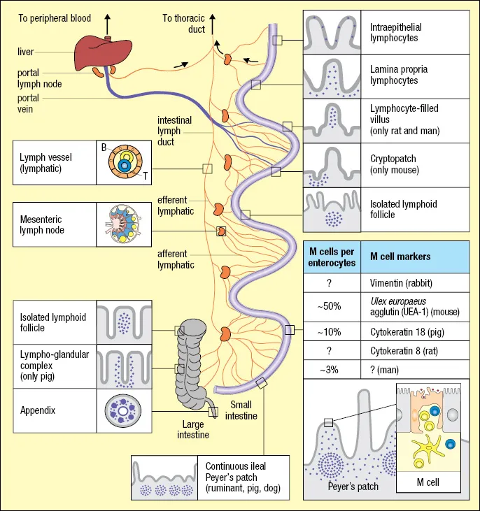
Figure 1.1 Distribution of lymphoid cells in various tissue compartments of the gut wall with species differences indicated. Lymphocytes leave the gut wall via draining lymphatics afferent to mesenteric lymph nodes, or via portal blood reaching the liver. Important regulation of immunity takes place in these organs, particularly induction of tolerance. The frequency of M cells in the follicle-associated epithelium of Peyer’s patches is highly variable, and a reliable marker for this specialized cell has not been identified in humans. In contrast to the antigen-dependent activation of B cells that takes place in Peyer’s patches of most species, the continuous ileal Peyer’s patch present in ruminants, pigs, and dogs appears to be a primary lymphoid organ responsible for antigen-independent B-cell development, similar to the bursa of Fabricius in chicken (not shown). This Peyer’s patch can be up to 2 meters long and constitute 80%–90% of the intestinal lymphoid tissue. (Adapted from Brandtzaeg, P., and Pabst, R. Trends Immunol. 2004, 25:570–577. With permission from Elsevier Ltd.)
The major component of human MALT comprises the gut-associated lymphoid tissue (GALT), including the PPs, the appendix, and numerous solitary follicles (see Figure 1.1) now termed “solitary isolated lymphoid tissue” (SILT) (Herbrand et al. 2008). Induction of mucosal immune responses can also take place in nasopharynx-associated lymphoid tissue (NALT), the tonsils (pharyngeal, palatine, lingual, pharyngeal), and bronchus-associated lymphoid tissue (BALT) as described later. Moreover, small MALT-like lymphoid aggregates are present in the conjunctiva and are associated with the larynx and various ducts such as those connecting the ocular and nasal compartments.
MALT resembles lymph nodes with B-cell follicles, interfollicular T-cell areas, and a variety of antigen-presenting cells (APCs) but lacks afferent lymphatics and a capsule. MALT therefore samples exogenous antigens directly from the mucosal surfaces through a follicle-associated epithelium (FAE), which contains “membrane” or “microfold” (M) cells (see Chapter 13). These specialized thin epithelial cells do not act as APCs but are effective in the uptake of microorganisms and other particulate antigens; they are also vulnerable “gaps” in the mucosal barrier. Studies in the mouse have, in addition, shown that dendritic cells (DCs) in the dome region of MALT send processes through the FAE to sample gut antigens.
The distinction between mucosal inductive and effector sites is not absolute, as the signals for extravasation and accumulation of naive versus memory/effector B and T cells are different. It is therefore confusing when MALT is used to refer to the mucosal effector compartments (e.g., the lamina propria and the surface epithelium of the gut and its diffusely distributed immune cells, Figure 1.2). This is in conflict with the classical definition of a lymphoid tissue, and so the Society for Mucosal Immunology and the International Union of Immunological Societies have agr...