
eBook - ePub
Musculoskeletal MRI E-Book
Nancy M. Major, Mark W. Anderson
This is a test
Compartir libro
- 480 páginas
- English
- ePUB (apto para móviles)
- Disponible en iOS y Android
eBook - ePub
Musculoskeletal MRI E-Book
Nancy M. Major, Mark W. Anderson
Detalles del libro
Vista previa del libro
Índice
Citas
Información del libro
Ideal for residents, practicing radiologists, and fellows alike, this updated reference offers easy-to-understand guidance on how to approach musculoskeletal MRI and recognize abnormalities. Concise, to-the-point text covers MRI for the entire musculoskeletal system, presented in a highly templated format. Thoroughly revised and enhanced with full-color artwork throughout, this resource provides just the information you need to perform and interpret quality musculoskeletal MRI.
- Includes the latest protocols, practical advice, tips, and pearls for diagnosing conditions impacting the temporomandibular joint, shoulder, elbow, wrist/hand, spine, hips and pelvis, knee, and foot and ankle.
- Follows a quick-reference format throughout, beginning with basic technical information on how to obtain a quality examination, followed by a discussion of the normal appearance and the abnormal appearance for each small unit that composes a joint.
- Depicts both normal and abnormal anatomy, as well as disease progression, through more than 600 detailed, high-quality images, most of which are new to this edition.
- Features key information boxes throughout for a quick review of pertinent material.
Preguntas frecuentes
¿Cómo cancelo mi suscripción?
¿Cómo descargo los libros?
Por el momento, todos nuestros libros ePub adaptables a dispositivos móviles se pueden descargar a través de la aplicación. La mayor parte de nuestros PDF también se puede descargar y ya estamos trabajando para que el resto también sea descargable. Obtén más información aquí.
¿En qué se diferencian los planes de precios?
Ambos planes te permiten acceder por completo a la biblioteca y a todas las funciones de Perlego. Las únicas diferencias son el precio y el período de suscripción: con el plan anual ahorrarás en torno a un 30 % en comparación con 12 meses de un plan mensual.
¿Qué es Perlego?
Somos un servicio de suscripción de libros de texto en línea que te permite acceder a toda una biblioteca en línea por menos de lo que cuesta un libro al mes. Con más de un millón de libros sobre más de 1000 categorías, ¡tenemos todo lo que necesitas! Obtén más información aquí.
¿Perlego ofrece la función de texto a voz?
Busca el símbolo de lectura en voz alta en tu próximo libro para ver si puedes escucharlo. La herramienta de lectura en voz alta lee el texto en voz alta por ti, resaltando el texto a medida que se lee. Puedes pausarla, acelerarla y ralentizarla. Obtén más información aquí.
¿Es Musculoskeletal MRI E-Book un PDF/ePUB en línea?
Sí, puedes acceder a Musculoskeletal MRI E-Book de Nancy M. Major, Mark W. Anderson en formato PDF o ePUB, así como a otros libros populares de Medicine y Radiology, Radiotherapy & Nuclear Medicine. Tenemos más de un millón de libros disponibles en nuestro catálogo para que explores.
Información
1
Basic Principles of Musculoskeletal MRI
Although a detailed understanding of nuclear physics is not necessary to interpret magnetic resonance imaging (MRI) studies, it also is unacceptable to read passively whatever images you are given without concern for how the images are acquired or how they might be improved. Radiologists should have a solid understanding of the basic principles involved in acquiring excellent images. This chapter describes the various components that go into producing high-quality images, stressing the fundamental principles shared by all MRI scanners.
Every machine is different. Clinical scanners are now available at strengths ranging from 0.2 tesla (T) to 3.0T. Additionally, each vendor has its own language for describing its hardware, software, and scanning parameters, and an entire chapter could be devoted to deciphering the terms used by different manufacturers. Time spent learning the details of your machine with your technologists or physicists would be time well spent. If you are interested, read one of the excellent discussions of MRI physics in articles or other textbooks because, for the most part, in this book we leave the physics to the physicists.
What Makes a Good Image?
Lack of Motion
Motion is one of the greatest enemies of MRI (Fig. 1.1). It can arise from a variety of sources, such as cardiac motion, bowel peristalsis, and respiratory movement. For most musculoskeletal applications, motion usually stems from body movement related to patient discomfort. Patient comfort is of paramount importance because even if all the other imaging parameters are optimized, any movement would ruin the entire image.

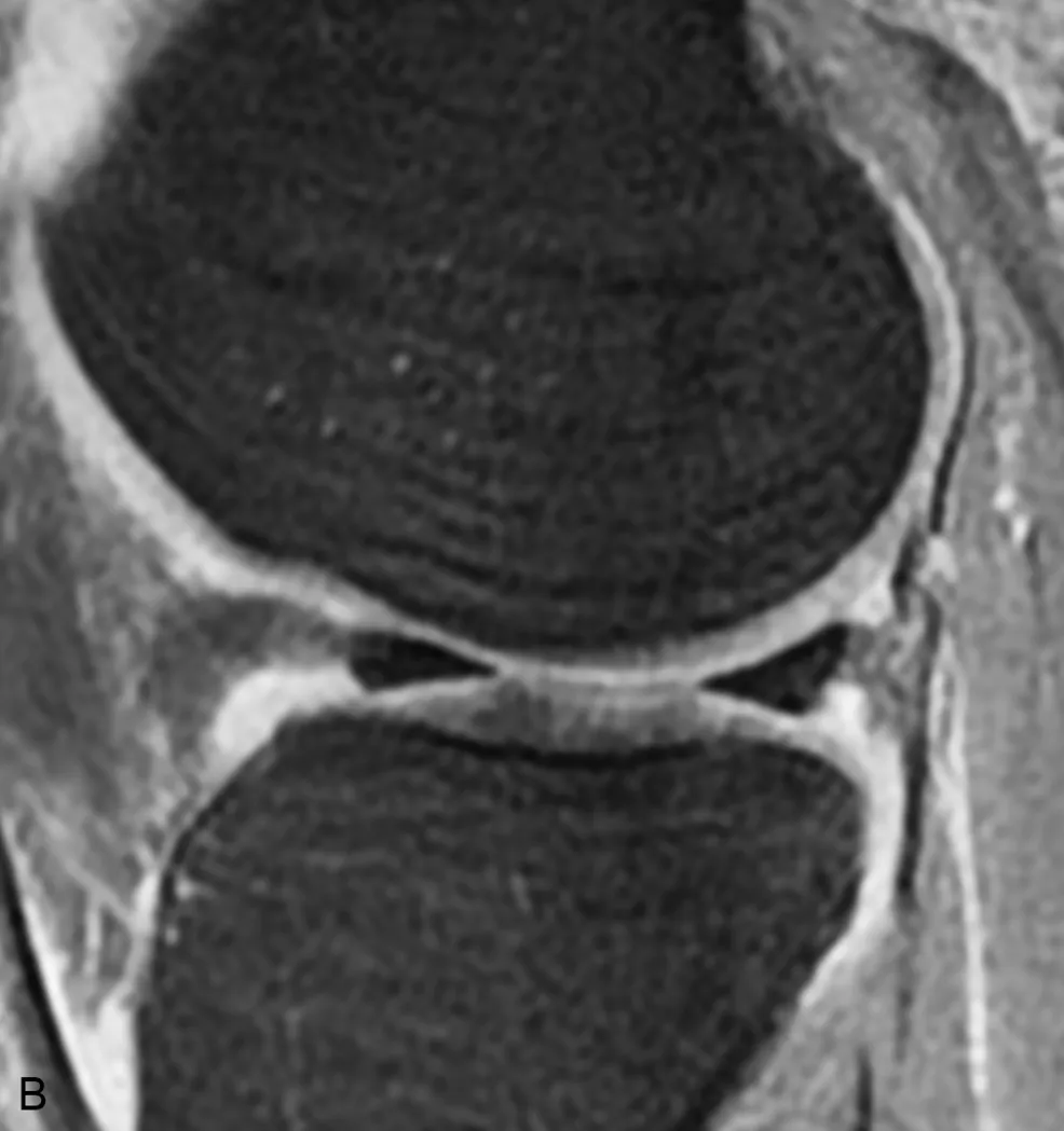
A, Sagittal proton density–weighted image of the knee. There is marked motion artifact and linear increased signal suggestive of a tear in the anterior horn of the lateral meniscus (arrowhead). B, Sagittal proton density with fat saturation is also degraded by motion artifacts but confirms that the meniscus is intact and that the meniscal signal abnormality was secondary to motion artifact.
Patient comfort begins with positioning. Every effort should be made to make the patient comfortable, such as placing a pillow beneath the knees when the patient is supine to reduce the stress on the back or providing padding at pressure points. When the patient is in a comfortable position, passive restraints, such as tape, foam rubber, or sandbags, can be used for maximal immobilization. Music via headphones can help alleviate anxiety. Short-acting sedation may be required for claustrophobic patients.
Another cause of patient motion is a prolonged examination, which is one reason why streamlined imaging protocols are useful. By designing efficient imaging sequences, the necessary scans are obtained in as short a time as possible, resulting in better patient compliance, improved technologist efficiency, and maximal scanner throughput. Standardized protocols also reduce the need for direct physician oversight during the scan and allow for improved image interpretation because the radiologist views the same anatomy in the same imaging planes utilizing the same sequences each time.
Signal and Resolution (Table 1.1)
Signal is the amount of information on an image. Other factors are important, but if the image is signal-poor (i.e., “noisy”), even the best radiologist would be unable to interpret it (Fig. 1.2).
Table 1.1
| ↑ Signal/↓ Resolution | ↑ Resolution/↓ Signal |
|---|---|
| ↑ Slice thickness | ↓ Slice thickness |
| ↑ Field of view | ↓ Field of view |
| ↓ Imaging matrix | ↑ Imaging matrix |

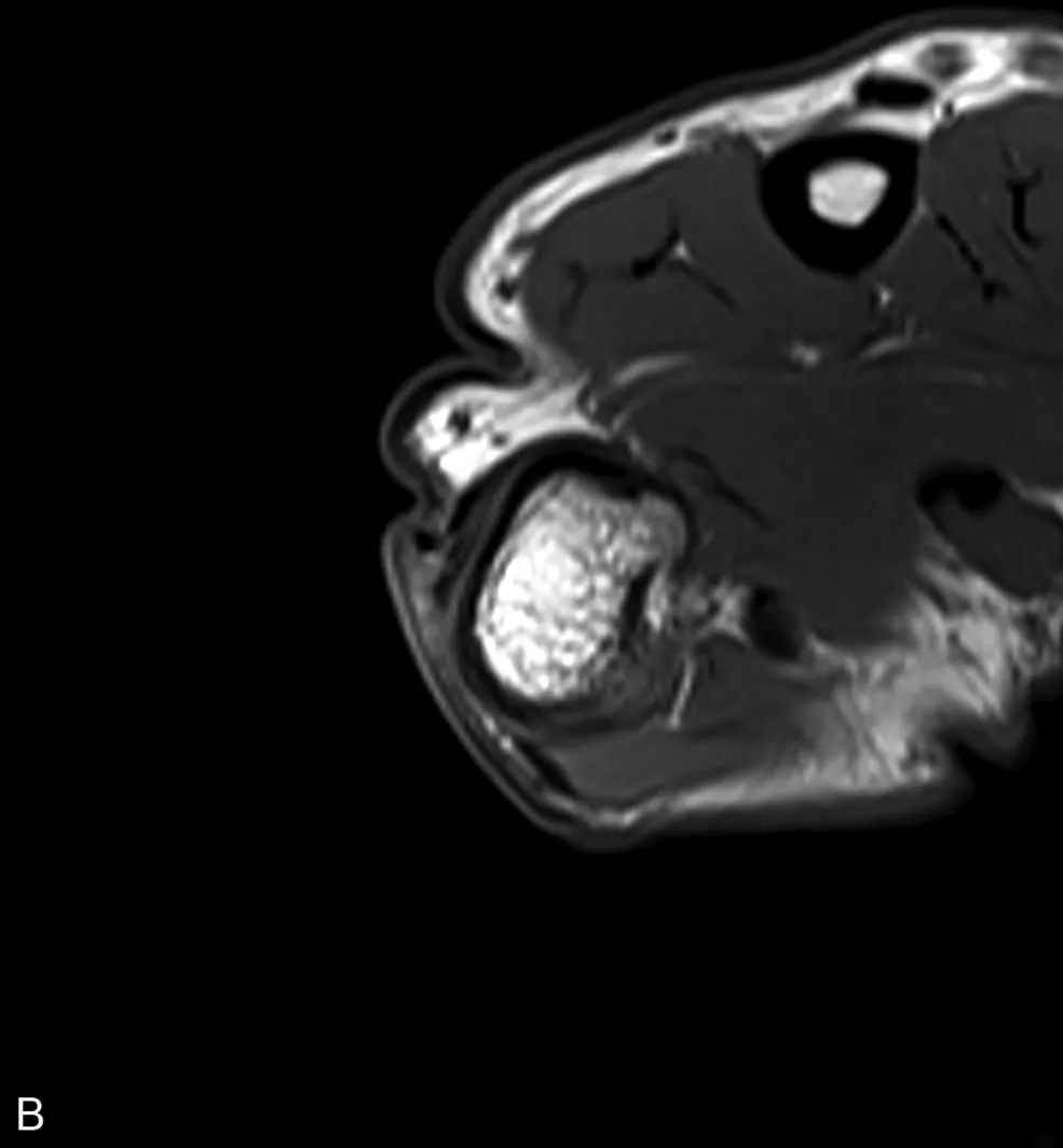
A, Axial T1-weighted image of the thumb obtained with a phased array extremity coil is of very poor quality, primarily related to prominent image noise. B, A follow-up axial T1-weighted image at the same level obtained with a dedicated wrist coil demonstrates markedly improved image quality due to an improved signal-to-noise ratio.
Each image is composed of voxels (volume elements) that correspond to small portions of tissue within the patient. One d...