
eBook - ePub
Musculoskeletal MRI E-Book
Nancy M. Major, Mark W. Anderson
This is a test
Buch teilen
- 480 Seiten
- English
- ePUB (handyfreundlich)
- Über iOS und Android verfügbar
eBook - ePub
Musculoskeletal MRI E-Book
Nancy M. Major, Mark W. Anderson
Angaben zum Buch
Buchvorschau
Inhaltsverzeichnis
Quellenangaben
Über dieses Buch
Ideal for residents, practicing radiologists, and fellows alike, this updated reference offers easy-to-understand guidance on how to approach musculoskeletal MRI and recognize abnormalities. Concise, to-the-point text covers MRI for the entire musculoskeletal system, presented in a highly templated format. Thoroughly revised and enhanced with full-color artwork throughout, this resource provides just the information you need to perform and interpret quality musculoskeletal MRI.
- Includes the latest protocols, practical advice, tips, and pearls for diagnosing conditions impacting the temporomandibular joint, shoulder, elbow, wrist/hand, spine, hips and pelvis, knee, and foot and ankle.
- Follows a quick-reference format throughout, beginning with basic technical information on how to obtain a quality examination, followed by a discussion of the normal appearance and the abnormal appearance for each small unit that composes a joint.
- Depicts both normal and abnormal anatomy, as well as disease progression, through more than 600 detailed, high-quality images, most of which are new to this edition.
- Features key information boxes throughout for a quick review of pertinent material.
Häufig gestellte Fragen
Wie kann ich mein Abo kündigen?
Gehe einfach zum Kontobereich in den Einstellungen und klicke auf „Abo kündigen“ – ganz einfach. Nachdem du gekündigt hast, bleibt deine Mitgliedschaft für den verbleibenden Abozeitraum, den du bereits bezahlt hast, aktiv. Mehr Informationen hier.
(Wie) Kann ich Bücher herunterladen?
Derzeit stehen all unsere auf Mobilgeräte reagierenden ePub-Bücher zum Download über die App zur Verfügung. Die meisten unserer PDFs stehen ebenfalls zum Download bereit; wir arbeiten daran, auch die übrigen PDFs zum Download anzubieten, bei denen dies aktuell noch nicht möglich ist. Weitere Informationen hier.
Welcher Unterschied besteht bei den Preisen zwischen den Aboplänen?
Mit beiden Aboplänen erhältst du vollen Zugang zur Bibliothek und allen Funktionen von Perlego. Die einzigen Unterschiede bestehen im Preis und dem Abozeitraum: Mit dem Jahresabo sparst du auf 12 Monate gerechnet im Vergleich zum Monatsabo rund 30 %.
Was ist Perlego?
Wir sind ein Online-Abodienst für Lehrbücher, bei dem du für weniger als den Preis eines einzelnen Buches pro Monat Zugang zu einer ganzen Online-Bibliothek erhältst. Mit über 1 Million Büchern zu über 1.000 verschiedenen Themen haben wir bestimmt alles, was du brauchst! Weitere Informationen hier.
Unterstützt Perlego Text-zu-Sprache?
Achte auf das Symbol zum Vorlesen in deinem nächsten Buch, um zu sehen, ob du es dir auch anhören kannst. Bei diesem Tool wird dir Text laut vorgelesen, wobei der Text beim Vorlesen auch grafisch hervorgehoben wird. Du kannst das Vorlesen jederzeit anhalten, beschleunigen und verlangsamen. Weitere Informationen hier.
Ist Musculoskeletal MRI E-Book als Online-PDF/ePub verfügbar?
Ja, du hast Zugang zu Musculoskeletal MRI E-Book von Nancy M. Major, Mark W. Anderson im PDF- und/oder ePub-Format sowie zu anderen beliebten Büchern aus Medicine & Radiology, Radiotherapy & Nuclear Medicine. Aus unserem Katalog stehen dir über 1 Million Bücher zur Verfügung.
Information
1
Basic Principles of Musculoskeletal MRI
Although a detailed understanding of nuclear physics is not necessary to interpret magnetic resonance imaging (MRI) studies, it also is unacceptable to read passively whatever images you are given without concern for how the images are acquired or how they might be improved. Radiologists should have a solid understanding of the basic principles involved in acquiring excellent images. This chapter describes the various components that go into producing high-quality images, stressing the fundamental principles shared by all MRI scanners.
Every machine is different. Clinical scanners are now available at strengths ranging from 0.2 tesla (T) to 3.0T. Additionally, each vendor has its own language for describing its hardware, software, and scanning parameters, and an entire chapter could be devoted to deciphering the terms used by different manufacturers. Time spent learning the details of your machine with your technologists or physicists would be time well spent. If you are interested, read one of the excellent discussions of MRI physics in articles or other textbooks because, for the most part, in this book we leave the physics to the physicists.
What Makes a Good Image?
Lack of Motion
Motion is one of the greatest enemies of MRI (Fig. 1.1). It can arise from a variety of sources, such as cardiac motion, bowel peristalsis, and respiratory movement. For most musculoskeletal applications, motion usually stems from body movement related to patient discomfort. Patient comfort is of paramount importance because even if all the other imaging parameters are optimized, any movement would ruin the entire image.

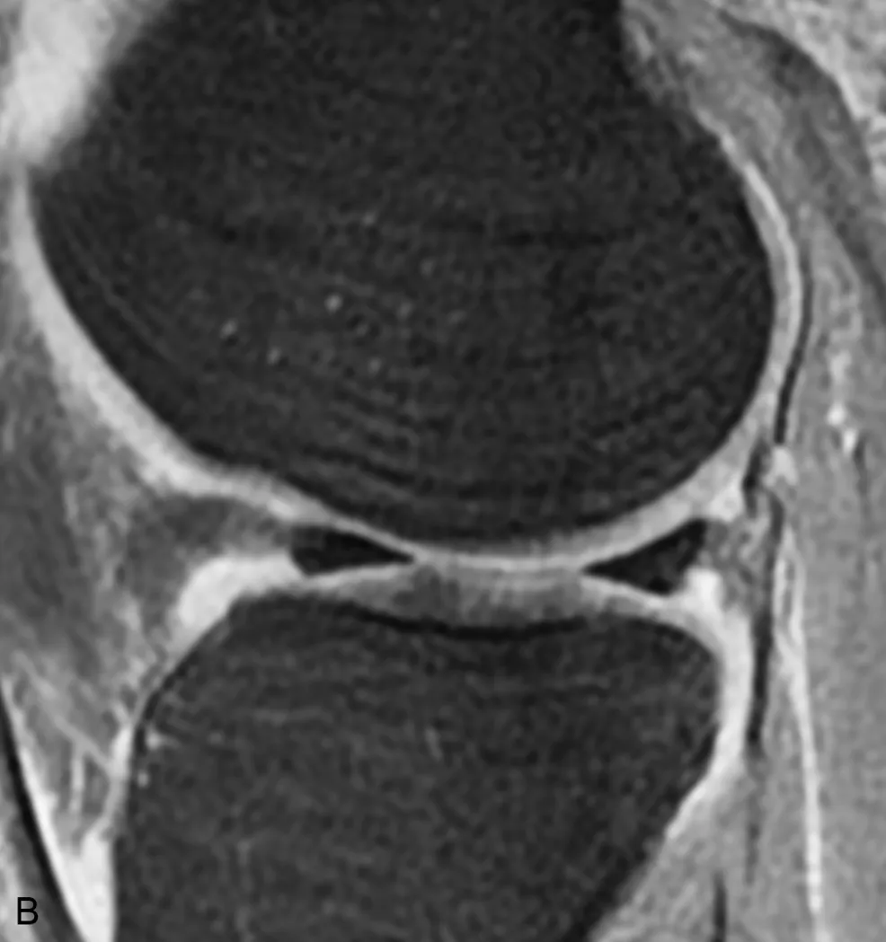
A, Sagittal proton density–weighted image of the knee. There is marked motion artifact and linear increased signal suggestive of a tear in the anterior horn of the lateral meniscus (arrowhead). B, Sagittal proton density with fat saturation is also degraded by motion artifacts but confirms that the meniscus is intact and that the meniscal signal abnormality was secondary to motion artifact.
Patient comfort begins with positioning. Every effort should be made to make the patient comfortable, such as placing a pillow beneath the knees when the patient is supine to reduce the stress on the back or providing padding at pressure points. When the patient is in a comfortable position, passive restraints, such as tape, foam rubber, or sandbags, can be used for maximal immobilization. Music via headphones can help alleviate anxiety. Short-acting sedation may be required for claustrophobic patients.
Another cause of patient motion is a prolonged examination, which is one reason why streamlined imaging protocols are useful. By designing efficient imaging sequences, the necessary scans are obtained in as short a time as possible, resulting in better patient compliance, improved technologist efficiency, and maximal scanner throughput. Standardized protocols also reduce the need for direct physician oversight during the scan and allow for improved image interpretation because the radiologist views the same anatomy in the same imaging planes utilizing the same sequences each time.
Signal and Resolution (Table 1.1)
Signal is the amount of information on an image. Other factors are important, but if the image is signal-poor (i.e., “noisy”), even the best radiologist would be unable to interpret it (Fig. 1.2).
Table 1.1
| ↑ Signal/↓ Resolution | ↑ Resolution/↓ Signal |
|---|---|
| ↑ Slice thickness | ↓ Slice thickness |
| ↑ Field of view | ↓ Field of view |
| ↓ Imaging matrix | ↑ Imaging matrix |

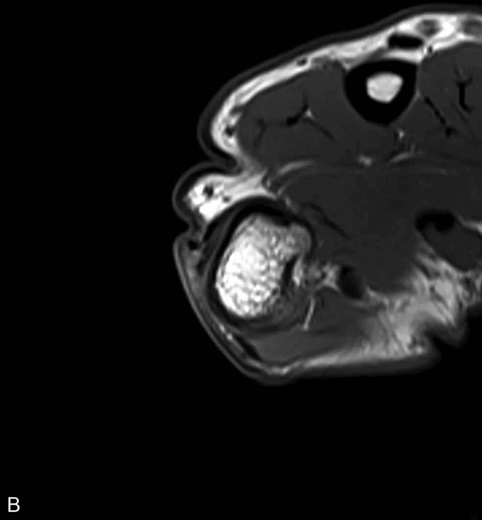
A, Axial T1-weighted image of the thumb obtained with a phased array extremity coil is of very poor quality, primarily related to prominent image noise. B, A follow-up axial T1-weighted image at the same level obtained with a dedicated wrist coil demonstrates markedly improved image quality due to an improved signal-to-noise ratio.
Each image is composed of voxels (volume elements) that correspond to small portions of tissue within the patient. One d...