![]()
Different Colon Diseases with Chronic Constipation
B.1
Pathogenesis of Hirschsprung’s Disease
Neuroblasts of the neck vagus migrate during embryonic weeks 6-12 craniocaudally along circular muscles to form a myenteric plexus. With a delay of several days, nerve cells of the myenteric plexus migrate into the submucosa to generate a submucous plexus.
During embryonic weeks 5-12, nerve fibers invade into circular muscles and mucosa of the distal colon from the sacral roots S2-S5. This happens 4 to 5 weeks before neuroblasts arrive in the descending colon. These nerves from the sacrum release acetylcholine in a synchronous manner. This causes a spastic contraction in the rectosigmoid up to the synaptic linking of these nerves with the invading neurocrest cells. This is a long-lasting process from embryonic weeks 10-12. It is a very sensitive period during which neuroblasts connect synaptic with parasympathetic nerves of sacral roots S2-S5. If neuroblasts do not arrive in the distal colon, HD or an aganglionosis develops. Therefore, HD is limited to the rectosigmoid or rectum in about 75% of babies.
In cases of rectum aganglionosis, nerve cells which modulate the permanent firing nerves from the sacral roots are missing, causing a spastic contraction of circular muscles and thus gut obstruction. Because acetylcholine and AChE have the same level, AChE can be used to
evaluate the cholinergic level of a particular tissue. This fact is used in enzyme histochemistry to recognize an aganglionosis in the rectal mucosa. The increase of AChE activity in parasympathetic nerve fibers of the rectum mucosa can be used as a reliable indicator of an aganglionosis. AChE activity increases dependently with age (fig. 2-6). LDH and SDH or nitroxide synthase (NOS), which stain nerve cells electively, can be used as a second indicator of an aganglionosis (fig. 7).
Indicators of Hirschsprung’s Disease
1 Increased acetylcholine release and AChE activity of parasympathetic nerves in lamina propria mucosae, muscularis mucosae, and muscularis propria are the result of an aganglionosis of the submucous and myenteric plexus (fig. 2-6).
2 A permanent release of acetylcholine by an inborn aganglionosis causes a spasticity of the muscularis propria in the distal colon.
3 The aganglionic segment of the rectum or rectosigmoid causes an obstruction in the distal colon.
Immaturity of nerve cells, often observed in young babies, show low SDH and LDH activity in nerve cells. In these cases, an unspecific reaction of NOS or NADH diaphorase may be helpful to establish aganglionosis of the submucosa. It is a great advantage to use only mucosa biopsies for HD diagnosis, thus avoiding anesthesia for a whole-mount biopsy or a laparoscopic seromuscular biopsy of the sigmoid [4, 13, 20, 23, 24].
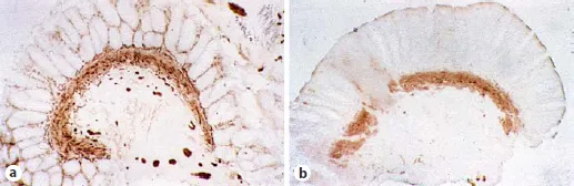
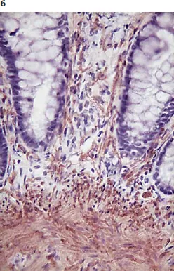
Fig. 2. a HD. Characteristic increase of AChE in parasympathetic nerve fibers of lamina propria mucosae and muscularis mucosae (2-week-old boy; compare with fig. 97). b Normal innervated rectum mucosa; 4-week-old boy. AChE staining. × 45.
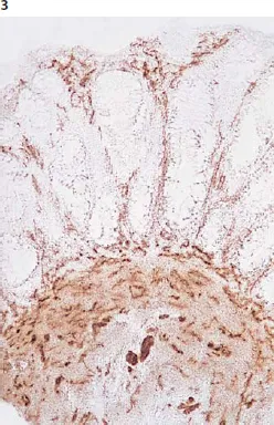
Fig. 3. Mucosa biopsy from the rectum (native cryostat section) with characteristic increase of AChE activity in parasympathetic nerve fibers in muscularis mucosae and lamina propria mucosae: HD. AChE reaction without counterstaining. × 90.
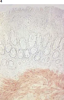
Fig. 4. AChE reaction of normal innervated rectum mucosa (native cryostat section). Only in muscularis mucosae are weakly stained nerve fibers to be observed. × 90.
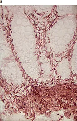
Fig. 5. Higher magnification of aganglionic mucosa biopsy section without counterstaining (7-month-old boy). × 180.
Fig. 6. Aganglionic rectum mucosa as in figure 5 with hemalum counterstaining. The identification of increased AChE activity in nerve fibers is much more difficult than in figure 5 without counterstaining. × 180.
In order not to miss a distal aganglionosis, the first biopsy may be taken from the rectoanal transition. Biopsies which are taken proximal from the anal ring may be taken in geometric distances (2, 4, and 8 cm above the anal ring). A seromuscular biopsy of the sigmoid allows no exclusion of an aganglionosis in the distal rectum [25, 26].
An enzyme histochemical AChE reaction in native rectal...