
eBook - ePub
A Beginner's Guide to Blood Cells
Barbara J. Bain
This is a test
Compartir libro
- English
- ePUB (apto para móviles)
- Disponible en iOS y Android
eBook - ePub
A Beginner's Guide to Blood Cells
Barbara J. Bain
Detalles del libro
Vista previa del libro
Índice
Citas
Información del libro
The third edition of this popular pocket book, A Beginner's Guide to Blood Cells written by Professor Barbara Bain, provides a concise introduction to normal and abnormal blood cells and blood counts for trainees in haematology.
- Includes a brand new chapter on emergency morphology, designed to make the clinical significance and urgency of certain laboratory findings clear for biomedical scientists and to assist trainee haematologists in the recognition of major clinically important abnormalities
- Contains exceptional full colour images throughout
- Introduces important basic concepts of hematology, setting haematological findings in a clinical context
- Provides a fully updated self-assessment section
- An essential resource for trainee haematologists, biomedical scientists, and biomedical science and medical students
Preguntas frecuentes
¿Cómo cancelo mi suscripción?
¿Cómo descargo los libros?
Por el momento, todos nuestros libros ePub adaptables a dispositivos móviles se pueden descargar a través de la aplicación. La mayor parte de nuestros PDF también se puede descargar y ya estamos trabajando para que el resto también sea descargable. Obtén más información aquí.
¿En qué se diferencian los planes de precios?
Ambos planes te permiten acceder por completo a la biblioteca y a todas las funciones de Perlego. Las únicas diferencias son el precio y el período de suscripción: con el plan anual ahorrarás en torno a un 30 % en comparación con 12 meses de un plan mensual.
¿Qué es Perlego?
Somos un servicio de suscripción de libros de texto en línea que te permite acceder a toda una biblioteca en línea por menos de lo que cuesta un libro al mes. Con más de un millón de libros sobre más de 1000 categorías, ¡tenemos todo lo que necesitas! Obtén más información aquí.
¿Perlego ofrece la función de texto a voz?
Busca el símbolo de lectura en voz alta en tu próximo libro para ver si puedes escucharlo. La herramienta de lectura en voz alta lee el texto en voz alta por ti, resaltando el texto a medida que se lee. Puedes pausarla, acelerarla y ralentizarla. Obtén más información aquí.
¿Es A Beginner's Guide to Blood Cells un PDF/ePUB en línea?
Sí, puedes acceder a A Beginner's Guide to Blood Cells de Barbara J. Bain en formato PDF o ePUB, así como a otros libros populares de Medizin y Hämatologie. Tenemos más de un millón de libros disponibles en nuestro catálogo para que explores.
Información
CHAPTER 1
The Blood Film and Count
Blood
Blood is a life‐sustaining fluid that circulates through the heart and blood vessels. It carries oxygen and nutrients to the tissues and waste products to the lungs, liver and kidneys, where they can be removed from the body. Usually when blood is removed from the body it forms a solid blood clot. However, if clotting is prevented by mixing with an anticoagulant, the blood separates, under the influence of gravity, into three layers (Fig. 1.1). The bottom layer is deep red in colour and is composed of red cells. The top layer is clear and pale yellow. It is called plasma and is composed of various salts and proteins dissolved in water. In between is a narrow layer called the buffy coat because of its buff or yellowish white colour. The buffy coat is composed mainly of cells of a variety of types, collectively known as white cells. In addition there are small cellular fragments, called platelets, which have a role in blood clotting.
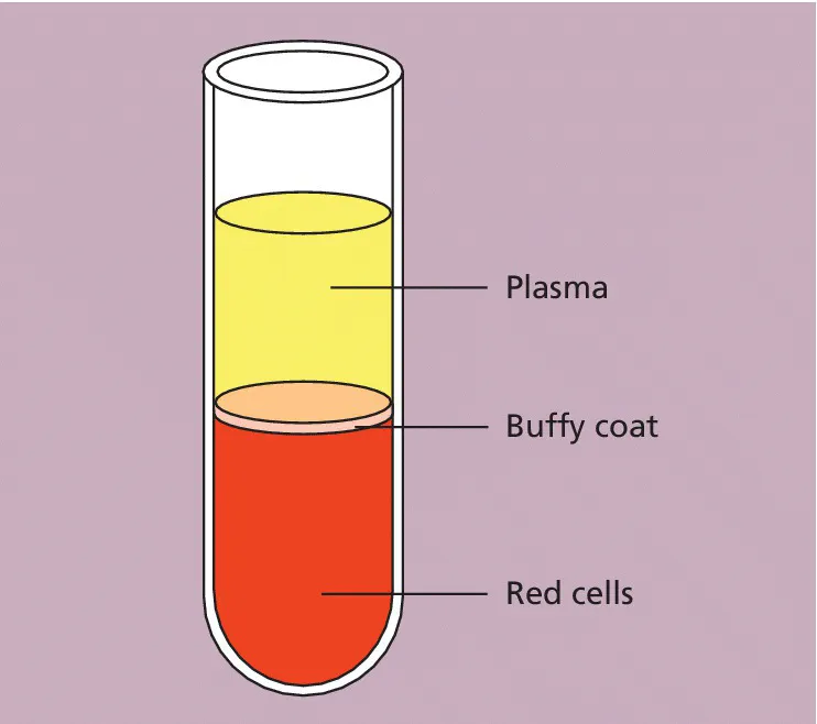
Fig. 1.1 Diagram of a tube of anticoagulated blood that has been allowed to sediment, showing the separation of blood into red cells, a buffy coat (white cells and platelets) and plasma.
The blood film
Although we can judge the proportions of red cells and white cells in a tube of sedimented blood, we get far more information if the blood is carefully mixed and a thin layer is spread on a glass slide to form a blood film. The blood cells are then preserved by exposure to the alcohol methanol, a process known as fixation. The fixed film of blood is stained with a mixture of several dyes so that the individual cells can be recognized when they are examined with a microscope. After staining, the colour of red cells is enhanced and the white cells and platelets, which would otherwise be transparent and colourless, have acquired a variety of colours that allow their detailed structure to be recognized. One of the commonest mixtures of dyes used to stain blood cells is the May–Grünwald–Giemsa (MGG) stain, named after its inventors. All the photographs in this book are of MGG‐stained blood films.
Red cells
The most numerous cells in a blood film are the red cells, also known as erythrocytes. Normal red cells are disc‐shaped but are thinner in the centre (Fig. 1.2). As a consequence, on a stained blood film, they have a circular outline and a paler central area (Fig. 1.3). Red cells owe their pinkish‐brown colour to the presence of a complex protein, haemoglobin, which is their major constituent. Enhancement of their colour in a stained film is because haemoglobin takes up eosin, one of the dyes of the MGG stain. In the body it is haemoglobin of the red cells that, in the lungs, combines with oxygen from inspired air and transports it to tissues where it is needed for the metabolic processes supplying the energy needs of the body. Mature red cells in humans (although not in some other species) differ from most body cells in that they do not have a nucleus. Red cells are produced in the bone marrow and usually lose their nuclei when they are released into the blood stream.

Fig. 1.2 A diagram of a red cell viewed from above and in cross‐section.
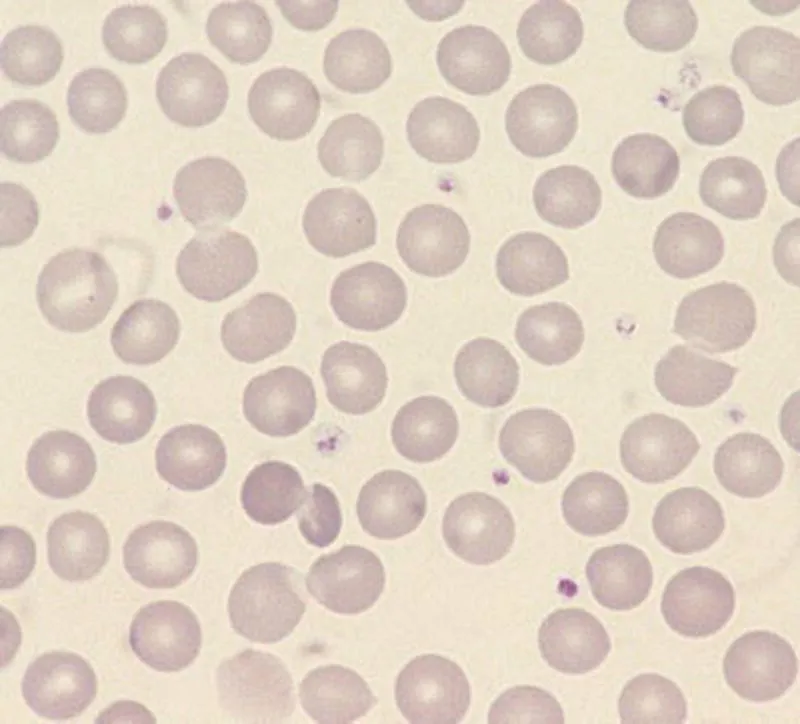
Fig. 1.3 Normal red cells (erythrocytes) showing little variation in size and shape, an approximately round outline and a small area of central pallor in some of the cells. The small structures containing lilac‐staining granules between the red cells are platelets.
White cells
In healthy people there are at least five types of white cell, or leucocyte, in the circulating blood. Unlike red cells, white cells have retained their nuclei. The cell is therefore made up of a nucleus and cytoplasm. The cytoplasm is the site of protein synthesis and other cellular functions. The nucleus is composed of chromatin, which is mainly deoxyribonucleic acid (DNA), carrying genetic messages. Genetic messages are transmit...