Samuel Webster, Geraint Morris, Euan Kevelighan, Samuel Webster, Geraint Morris, Euan Kevelighan
This is a test
This is a test
Compartir libro
English
ePUB (apto para móviles)
Disponible en iOS y Android
eBook - ePub
Essential Human Development
Samuel Webster, Geraint Morris, Euan Kevelighan, Samuel Webster, Geraint Morris, Euan Kevelighan
Detalles del libro
Vista previa del libro
Índice
Citas
Información del libro
As our understanding of the human body broadens, so does the need for a comprehensive text that encompasses all aspects of human development. Essential Human Development is a great course companion that focuses on the human life cycle, ideal for the undergraduate student new to these fields, or for qualified practitioners looking for a reference guide.
Featuring key information points and self-test assessments in each chapter, the book is organised in an accessible manner, beginning with fertilisation and embryology, then moving on to obstetric medicine, neonatal care and child health, with the final section exploring gynaecological medicine.
Ensuring that information is placed in context to aid understanding, Essential Human Development is the perfect support for the modern medical school curriculum, as well as a vital reminder of the core information needed whilst on a women or child health clinical placement.
Preguntas frecuentes
¿Cómo cancelo mi suscripción?
Simplemente, dirígete a la sección ajustes de la cuenta y haz clic en «Cancelar suscripción». Así de sencillo. Después de cancelar tu suscripción, esta permanecerá activa el tiempo restante que hayas pagado. Obtén más información aquí.
¿Cómo descargo los libros?
Por el momento, todos nuestros libros ePub adaptables a dispositivos móviles se pueden descargar a través de la aplicación. La mayor parte de nuestros PDF también se puede descargar y ya estamos trabajando para que el resto también sea descargable. Obtén más información aquí.
¿En qué se diferencian los planes de precios?
Ambos planes te permiten acceder por completo a la biblioteca y a todas las funciones de Perlego. Las únicas diferencias son el precio y el período de suscripción: con el plan anual ahorrarás en torno a un 30 % en comparación con 12 meses de un plan mensual.
¿Qué es Perlego?
Somos un servicio de suscripción de libros de texto en línea que te permite acceder a toda una biblioteca en línea por menos de lo que cuesta un libro al mes. Con más de un millón de libros sobre más de 1000 categorías, ¡tenemos todo lo que necesitas! Obtén más información aquí.
¿Perlego ofrece la función de texto a voz?
Busca el símbolo de lectura en voz alta en tu próximo libro para ver si puedes escucharlo. La herramienta de lectura en voz alta lee el texto en voz alta por ti, resaltando el texto a medida que se lee. Puedes pausarla, acelerarla y ralentizarla. Obtén más información aquí.
¿Es Essential Human Development un PDF/ePUB en línea?
Sí, puedes acceder a Essential Human Development de Samuel Webster, Geraint Morris, Euan Kevelighan, Samuel Webster, Geraint Morris, Euan Kevelighan en formato PDF o ePUB, así como a otros libros populares de Medicine y Gynecology, Obstetrics & Midwifery. Tenemos más de un millón de libros disponibles en nuestro catálogo para que explores.
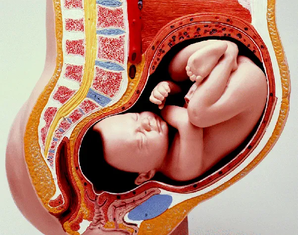
Jamie is a 4-month-old boy presenting with disparity between limb length, trunk length and cranial circumference. His height is under the fourth percentile, his weight is under the fourth percentile and his head circumference is above the 97th percentile. Motor development milestones are delayed. Jamie's mother and father have typical heights (168 cm and 176 cm respectively).
Learning Outcomes
You should be able to recognise the stages of cell division in mitosis and meiosis.
You should be able to describe the basic principles of growth and differentiation.
Chromosomes
As a basis of biology cell theory is a crucial part of understanding development. Complex organisms grow from a single cell. The cell is the fundamental unit of structure in the organism, and new cells are formed from existing cells. All structure, function and organisation relates to the unit of the cell. In development we consider how the cells of the gametes merge to form a cell with a new genetic composition, the division of that cell to form new cells, and how those cells become organised, form shapes and tissues of multiple differentiated cell types.
DNA is stored in chromatin form within the nuclei of cells, and RNA is present in the cytoplasm. When cells divide the chromosomes are duplicated and the daughter cells gain exact copies of the DNA of the parent cell (hopefully, if the replication and error checking mechanisms work correctly).
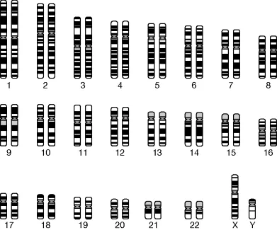
Somatic cells contain 23 pairs of chromosomes including 22 pairs of autosomes and one pair of sex chromosomes (Figure 1.1). Each chromosome is an organised package of DNA.
Figure 1.1Human karyotype. (Source: S. Webster and R. de Wreede (2016) Embryology at a Glance, 2nd edn. Reproduced with permission of John Wiley & Sons, Ltd.)
In a homologous pair of chromosomes the same genes are encoded on each chromosome but the genes may occur as slightly different versions. One chromosome has been inherited from the father, and the other from the mother. For example, the gene for head hair pigment colour will occur on both chromosomes of a homologous pair, but one copy may encode for blonde hair and the other for brown. These copies are alleles, and the dominant pigment allele will be represented in the phenotype of the individual. This is a simplified example, and many hair pigments are at play in determining a person's final hair colour, accounting for the wide variation of natural shades that occurs. The mixing up of alleles across homologous chromosomes during cell division is an important part of the genetic diversity advantage given by sexual reproduction over asexual reproduction.
If a cell has two copies of each kind of chromosome (e.g. one copy from the mother and one copy from the mother) it is said to be diploid. If it only had one copy it would be haploid.
We can also describe a cell by the number of copies (n) of each unique double-stranded length of chromosomal DNA. Chromosomal DNA inherited from the mother is different to chromosomal DNA inherited from the father. In a pair of chromosomes the genes are the same but the alleles are different. A haploid cell has only one copy of each kind of chromosome so it is described as 1n. Somatic cells are normally diploid, and during part of the cell cycle only have one DNA strand for each kind of chromosome so are described as 2n. They have two copies of each kind of chromosome (one from the mother and one from the father). When a cell copies its DNA in preparation for cell division it will have four copies of each kind of chromosome and be described as 4n.
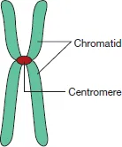
If the DNA strand of a chromosome is duplicated its two duplicates are joined together at the centromere forming the familiar X shape of most chromosomes (Figure 1.2). Each of the two duplicates is a sister chromatid.
Figure 1.2The structure of a chromosome. (Source: S. Webster and R. de Wreede (2016) Embryology at a Glance, 2nd edn. Reproduced with permission of John Wiley & Sons, Ltd.)
Mitosis
Mitosis is the process by which cells divide and increase in number in eukaryotic organisms. The result of mitosis is two daughter cells that contain the same genetic information. Mitosis is the method by which cells repair tissues, it is one way in which growth can occur, and it is how cells lost through normal processes are replaced. Some cells are very good at proliferating by mitosis, such as epidermal keratinocytes, which are lost daily as flakes of skin, and some cells are very poor at mitotic division, such as neurones of the central nervous system, which are expected to survive for the lifetime of the organism (although it is not yet clearly understood how long neurones live, but they are not naturally replaced after brain damage). Mitosis is a major mechanism of growth in the embryo and fetus.
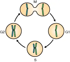
Cell division is a step within the cell cycle (Figure 1.3). The cell cycle describes a series of carefully controlled events in the life of cell that take part in cell division, and cells that do not divide are considered to have left the cell cycle. The stages of the cell cycle are gap 1 (G1), synthesis (S), gap 2 (G2) and mitosis (M). The stages of G1, S and G2 are also known collectively as interphase. A cell's DNA is duplicated during S phase, adding a sister chromatid to the existing chromatid. A cell that no longer divides can be described as existing within a G0 phase.
Figure 1.3The cell cycle. (Source: S. Webster and R. de Wreede (2016) Embryology at a Glance, 2nd edn. Reproduced with permission of John Wiley & Sons, Ltd.)
When a cell begins mitosis its chromosomes become condensed and form their recognisable X shapes during the first phase of mitosis, called prophase (Figure 1.4). At this stage it is diploid (4n). Centrioles are cylindrical structures that have a number of functions within eukaryotic cells, and during mitosis they arrange and separate DNA. During prophase the centrioles move to opposite ends of the cell.
Figure 1.4Mitosis. (Source: S. Webster and R. de Wreede (2016) Embryology at a Glance, 2nd edn. Reproduced with permission of John Wiley & Sons, Ltd.)
In the next stage, prometaphase, the nuclear membrane breaks down and disappears releasing the DNA into the cytoplasm. Microtubules link the centromeres of the chromosomes to the centrioles, and during metaphase the chromosomes begin to move, pulled by the microtubules to line up along the middle of the cell.
The centromeres are cut in the telophase step, splitting each chromosome into its separate, genetically identical chromatids. One of each pair of chromatids is pulled to opposite ends of the cell by microtubules and the centrioles.
In telophase the chromatids reach the ends of the cell, begin to lengthen again and are no longer visible under a light microscope. Two new nuclear membranes begin to form around the chromatid DNA to create two nuclei. Cytokinesis follows during which a ring of actin filaments appears around the midline of the cell and shrinks, splitting the cell into two. Mitosis is complete, and the two cells return to the G1 phase. During the G1 phase each cell has a full, diploid complement of DNA but only one copy of each chromosome (2n).