William J. Reagan, Armando R. Irizarry Rovira, Dennis B. DeNicola
This is a test
This is a test
Compartir libro
English
ePUB (apto para móviles)
Disponible en iOS y Android
eBook - ePub
Veterinary Hematology
Atlas of Common Domestic and Non-Domestic Species
William J. Reagan, Armando R. Irizarry Rovira, Dennis B. DeNicola
Detalles del libro
Vista previa del libro
Índice
Citas
Información del libro
Now in its third edition, Veterinary Hematology: Atlas of Common Domestic and Non-Domestic Species continues to offer veterinarians and veterinary technicians an essential guide to veterinary hematology. Comprehensive in scope, the atlas presents the fundamentals of both normal and abnormal blood cell morphologies, with coverage of a wide range of species, including dogs, cats, horses, ruminants, llamas, rats, mice, nonhuman primates, ferrets, rabbits, guinea pigs, birds, amphibians, and reptiles.
Designed as a useful and accessible guide, the updated third edition presents more than 300 color images and includes a new chapter that describes the best techniques for using hematology instruments.The authors—noted experts on the topic—clearly show how to identify and interpret the hematological changes that may occur in a variety of species. In addition, a companion website offers a wealth of additional hematological images. This vital atlas:
Provides an updated edition of the popular veterinary hematology atlas for veterinarians, veterinary students, and veterinary technicians
Contains a new instructive chapter on hematology instrumentation
Presents hundreds of high-quality color photographs that help in identification
Covers a range of species from dogs and cats to birds and reptiles
Features a companion website that provides a wealth of hematological images
Written for both novice and experienced veterinarians, Veterinary Hematology provides a complete resource to blood morphologic abnormalities in domestic and non-domestic species.
Preguntas frecuentes
¿Cómo cancelo mi suscripción?
Simplemente, dirígete a la sección ajustes de la cuenta y haz clic en «Cancelar suscripción». Así de sencillo. Después de cancelar tu suscripción, esta permanecerá activa el tiempo restante que hayas pagado. Obtén más información aquí.
¿Cómo descargo los libros?
Por el momento, todos nuestros libros ePub adaptables a dispositivos móviles se pueden descargar a través de la aplicación. La mayor parte de nuestros PDF también se puede descargar y ya estamos trabajando para que el resto también sea descargable. Obtén más información aquí.
¿En qué se diferencian los planes de precios?
Ambos planes te permiten acceder por completo a la biblioteca y a todas las funciones de Perlego. Las únicas diferencias son el precio y el período de suscripción: con el plan anual ahorrarás en torno a un 30 % en comparación con 12 meses de un plan mensual.
¿Qué es Perlego?
Somos un servicio de suscripción de libros de texto en línea que te permite acceder a toda una biblioteca en línea por menos de lo que cuesta un libro al mes. Con más de un millón de libros sobre más de 1000 categorías, ¡tenemos todo lo que necesitas! Obtén más información aquí.
¿Perlego ofrece la función de texto a voz?
Busca el símbolo de lectura en voz alta en tu próximo libro para ver si puedes escucharlo. La herramienta de lectura en voz alta lee el texto en voz alta por ti, resaltando el texto a medida que se lee. Puedes pausarla, acelerarla y ralentizarla. Obtén más información aquí.
¿Es Veterinary Hematology un PDF/ePUB en línea?
Sí, puedes acceder a Veterinary Hematology de William J. Reagan, Armando R. Irizarry Rovira, Dennis B. DeNicola en formato PDF o ePUB, así como a otros libros populares de Medicine y Veterinary Medicine. Tenemos más de un millón de libros disponibles en nuestro catálogo para que explores.
All blood cells have a finite life span, but in normal animals, the number of cells in circulation is maintained at a fairly constant level. To accomplish this, cells in circulation need to be constantly replenished, which occurs via the production and release of cells from the bone marrow. Production sites in the bone marrow are commonly referred to as medullary sites. In times of increased demand, production can also occur outside the bone marrow in sites such as spleen, liver, and lymph nodes. These sites are called extramedullary sites. In rodents, in the normal steady state, extramedullary production of blood cells occurs in the spleen.
Hematopoiesis, the production of blood cells, is a complex and highly regulated process. Some differences in hematopoiesis exist between species and are beyond the scope of this text; readers are referred to the detailed coverage in some of the references in Bibliography section. The dog will be used to demonstrate some of the basic principles of hematopoiesis. All blood cells in the bone marrow arise from a common stem cell. This pluripotent stem cell gives rise to several stages of committed progenitor cells, which then differentiate into cells of the erythrocytic, granulocytic, megakaryocytic, and agranulocytic (monocytic and lymphocytic) lineages. The end result of this development process is the release of red blood cells, white blood cells, and platelets into the circulation. At the light microscopic level, without the use of immunocytochemistry or enzyme cytochemistry, it is impossible to accurately identify the early stem cells in the bone marrow, but the more differentiated stages of development can be identified and are graphically depicted in Figure 1.1.
Figure 1.1 Overview of hematopoiesis.
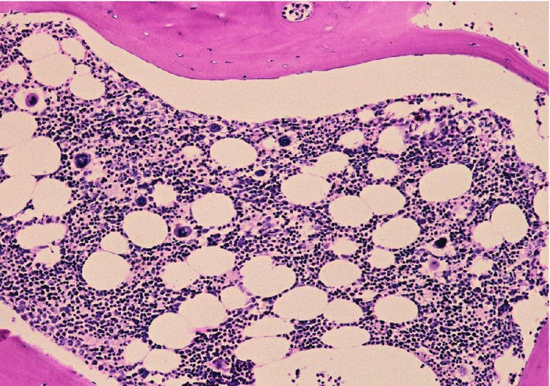
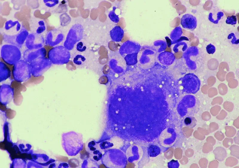
Figure 1.2 shows a histological section of a bone marrow core biopsy from an adult dog. Note that there is a mixture of approximately 50% hematopoietic cells and 50% fat that is surrounded by bony trabeculae. The specific types of bone marrow cells can be difficult to recognize in histological sections at this low-power magnification, but the very large cells present are megakaryocytes. Cells are easier to identify on a smear from a bone marrow aspirate (Figure 1.3). The cells that are present include erythrocytic and granulocytic precursors and a megakaryocyte. To classify these three different cell types, there are some general features that can be used. Megakaryocytes are easy to distinguish by their very large size; the majority of them are 100–200 μm in diameter compared with approximately 20–30 μm for the largest granulocytic or erythrocytic precursors.
Figure 1.2 Histological section of canine bone marrow. Pink bony trabeculae are present in the lower left corner, lower right corner, and top of the photomicrograph and surround the hematopoietic cells and fat. The round to oval clear areas are the fat. The erythrocytic and granulocytic precursor cells are the many small, round purple structures. The larger, densely staining purple structures distributed throughout the marrow space are megakaryocytes. Canine bone marrow core biopsy; hematoxylin and eosin stain; 10× objective.
Figure 1.3 Megakaryocyte, erythrocytic precursors, and granulocytic precursors. The megakaryocyte is the largest cell located in right center of the field. The early erythrocytic precursors have central round nuclei and deep blue cytoplasm. The early granulocytic precursors have oval to indented nuclei and blue cytoplasm. There is a granulocytic predominance in this field. Canine bone marrow smear; 50× objective.
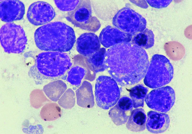
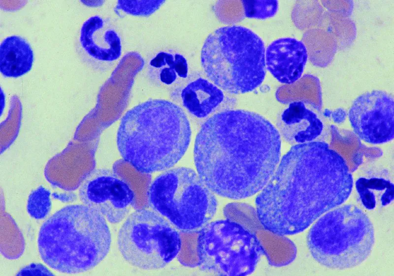
Cells of the erythrocytic lineage can be initially distinguished from those of the granulocytic lineage on the basis of their nuclear shape and color of cytoplasm (Figures 1.4 and 1.5). Cells of the erythrocytic lineage have very round nuclei throughout most stages of development. In contrast, the nuclei of cells of the granulocytic lineage become indented and segmented as they mature. In addition, the cytoplasm of early erythrocytic precursors is much bluer than that of the granulocytic precursors.
Figure 1.4 Erythrocytic precursors. The majority of the intact cells present are early erythrocytic precursors with centrally located round nuclei and deep blue cytoplasm. The cells with round eccentrically placed nuclei and reddish blue cytoplasm are late-stage erythrocytic precursors. The largest cell in the right center of the field that has small pink granules in the cytoplasm is a promyelocyte. Canine bone marrow smear; 100× objective.
Figure 1.5 Granulocytic precursors. The majority of the intact cells present are granulo...