![]()
1
INTRODUCTION
The kingdom Fungi consists of a distinct group of eukaryotic organisms that absorb their nourishment from living or dead organisms or organic matter. Fungi are found throughout nature, performing an essential service in returning to the soil nutrients removed by plants. There is, however, a large group of species that are parasitic on plants and a smaller group that are parasitic on animals, as well as on man. Fungi show considerable variation in size and form, but can be divided into three main groups: multicellular filamentous fungi (moulds); unicellular fungi (yeasts); and dimorphic fungi which are capable of changing their growth to either a multicellular or unicellular form, depending on the growth conditions.
In most multicellular fungi, the vegetative stage consists of a system of tubular, branching filaments, or mycelium. Each individual filament, or hypha, has a rigid cell wall and increases in length as a result of apical growth. In the more primitive fungi, the hyphae remain aseptate (without cross-walls). In the more advanced groups however, the hyphae are divided into compartments or cells by the development of more or less frequent cross-walls, termed septa. Such hyphae are termed septate.
Yeasts are unicellular fungi consisting of separate, round, oval or elongated cells or blastospores that propagate by an asexual process called budding in which the cell develops a protuberance from its surface. The bud enlarges and may become detached from the parent cell, or it may remain attached and itself produce another bud. In this way a chain of cells may be produced. Under certain conditions, continued elongation of the parent cell before it buds results in a chain of elongated cells, termed a pseudohypha, which resembles the hypha of moulds. Unlike a true hypha, however, the connection between adjacent pseudohyphal cells shows a marked constriction. Some yeasts can also produce true hyphae, with cross-walls. A small number of yeasts reproduce by fission. Yeasts are neither a natural nor a formal taxonomic group, but are a growth form shown by a wide range of unrelated fungi.
Some medically important fungi change their growth form during the process of tissue invasion. These dimorphic pathogens usually change from a multicellular hyphal form in the natural environment to a budding, single-celled yeast form in tissue.
Fungi reproduce by means of microscopic propagules, termed spores, that consist of a single cell or several cells contained within a rigid wall. Spores may be produced by an asexual process (involving mitosis only) or by sexual reproduction (involving meiosis). Some species of fungi are homothallic and able to form sexual structures within individual colonies. Most, however are heterothallic and do not form their sexual structures unless two different mating strains come into contact. Thus, sexual reproduction is often difficult to obtain in culture. The sexual spores and the structures in which they are produced form the traditional basis for fungal classification. Most recently the kingdom Fungi has been divided into a number of lesser groups, termed phyla, based on differences in their sexual structures. Two of these phyla (the Ascomycota and the Basidiomycota) and two sub-phyla (the Mucoromycotina and Entomophthoromycotina) contain species that are pathogenic to humans and animals.
Sexual Reproduction
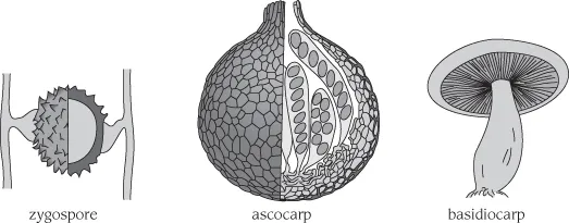
Of the kingdom Fungi, the majority of species belong to the sub-kingdom Dikarya (literally ‘two nuclei’ as their sexual reproduction involves a cell containing two fusing nuclei). This group is made up of two phyla (the Basidiomycota and the Ascomycota). Outside the Dikarya there are many other smaller groups, with sexual reproduction often involving the fusion of multiple nuclear pairs in a single cell. Examples of the latter are seen in the sub-phyla Mucoromycotina and Entomophthoromycotina. These two sub-phyla have replaced the phylum Zygomycota, a grouping now abandoned as misrepresenting phylogenetic relationships. Both show fusion of the multinucleate tips of two hyphae leading to the formation of a single, large zygospore, lying between them. This is a multinucleate thick-walled structure that has evolved to endure adverse environmental conditions. Meiosis occurs on germination and the vegetative haploid mycelium develops.
In contrast, in the Ascomycota and Basidiomycota, sexual reproduction has evolved into a means of rapid dispersal to new habitats, unlike the resting nature of the zygospore. In both these groups the diploid stage is transient, with meiosis resulting in the production of enormous numbers of short-lived haploid spores. In the Ascomycota, the sexual spores or ascospores are produced in sacs, or asci. Each ascus usually contains eight ascospores. The group shows a gradual transition from primitive forms that produce single asci to species that produce large structures, or ascocarps, containing large numbers of asci. Three main forms of ascocarp are common: the perithecium which releases its spores through an apical opening; the cleistothecium, which splits open to liberate its contents; and the gymnothecium, which is an open loose network of protective hyphae.
In most of the Basidiomycota the sexual spores or basidiospores are borne on projections at the tip of club-shaped basidia. These are produced in macroscopic structures or basidiocarps.
Whilst the reproductive structures associated with sexual life cycles are important to a full understanding of the fungi, most of the organisms described in this manual may be identified on the basis of their asexual reproductive structures and spores.
Asexual Reproduction
Fungi may also produce asexual spores by simple haploid nuclear division. Again, short lived propagules are produced in enormous numbers to ensure spread to new habitats. In many fungi this asexual (anamorph or imperfect) stage has proved so successful that the sexual (teleomorph or perfect) stage has diminished or even disappeared. These species have long been known as the Fungi Imperfecti or Deuteromycetes. This by convention contained all the asexual relatives of the Ascomycota and Basidiomycota, but not those of the former Zygomycota. With advances in molecular phylogenetic analysis, the concept of Fungi Imperfecti is becoming increasingly redundant as a useful taxonomic grouping, since most asexually-reproducing species can now be placed with their sexually reproducing relatives.
Conidia
In the Ascomycota and Basidiomycota the asexual spores are termed conidia, and are produced from a conidiogenous cell. In some species the conidiogenous cell is not different from the rest of the mycelium. In others the conidiogenous cell is contained in a specialised hyphal structure or conidiophore. There are two basic methods of asexual spore production: thallic in which an existing hyphal cell is converted into a conidium; and blastic, in which the conidium is produced as a result of some form of budding process.
Thallic Conidiogenesis
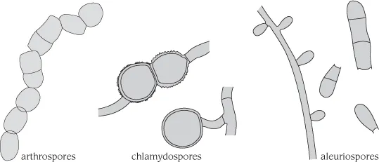
In thallic conidiogenesis the conidium is produced from an existing hyphal cell. This occurs when a hypha breaks up into sections to form individual cells, or arthrospores, or when one cell develops a thick wall to form a resting spore or chlamydospore.
Arthrospores are derived from the fragmentation of an existing hypha and represent the simplest form of asexual sporulation. In most species the septum separating two cells splits down the middle, leaving a trace of the resulting torn wall on the end of the spore. In a few instances the arthospores are intercalated with separating cells and are liberated after these cells have dissolved. This leaves a marked annular frill at the ends of the detached arthrospores. Moulds which produce arthrospores as their principal reproductive spores are described in detail in Chapter 3.
Aleuriospores represent an intermediate state between thallic and blastic conidiogenesis. These spores are formed from the side or tip of a hypha and during the initial stage before a septum is laid down, can resemble short, hyphal branches. As in all genuine cases of thallic conidiogenesis, it is not poss...