![]()
CHAPTER ONE
Preparation of Gametes
for Embryonic Development
THE CELL
The study of embryonic development requires an understanding not only of how and when structures arise and develop but also of how they enlarge and grow. The human individual arises from the conjugation of two minute structures called cells, one from the mother (oocyte) and one from the father (spermatozoon). These are called gametes. Together, these gametes form a single cell, the zygote, from which the entire embryo, including its surrounding membranes, grows. The zygote undergoes successive cleavages and the cells thus produced multiply so that all the organs of the embryo are developed. Thereafter, the formed organs grow and enlarge in the fetus until the moment of birth. The newborn grows into the infant, the adolescent and the adult. Cell growth and cell multiplication, therefore, are the most fundamental aspects of embryonic development and of existence, and are present from ‘conception’ to the ‘cessation’ of life.
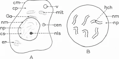
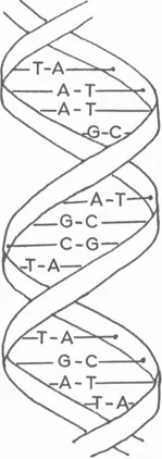
Before considering the actual process of cell multiplication or cell division, let us consider briefly those structures of a cell which are intimately associated with the process of cell division. The cell is regarded as the basic ‘working unit’ of all animals and plants. It is a minute living structure consisting of a mass of cytoplasm surrounded by a cell membrane. The cytoplasm contains a number of organelles and a nucleus (Fig. 1.1). The nucleus is also confined within a nuclear membrane and contains chromatin, a chemical substance composed of deoxyribonucleic acid (DNA). This substance consists of simple proteins (histones) combined with the bases adenine, thymine, guanine and cytosine. These are bound randomly in pairs (A-T, G-C etc.) across two complementary strands of DNA set in the form of a double helix (Fig. 1.2). Combinations of these base pairs form genes which confer upon individuals their distinguishing characteristics. The chromatin is condensed into chromosomes which, when suitably stained, is seen as strands under the microscope. Although much emphasis is placed upon the activity of the nucleus in cell multiplication, the cytoplasmic contents also play an important role in the process. The mitochondria provide the energy for the division, while the presence and splitting of the centrosome is essential for the process of division.
Figure 1.1: A diagram to illustrate the structure of a cell (A) and the formation of chromosomes in the nucleus (B). cen = centrosome; cm = cell membrane; cp = cytoplasm; cs = chromatic substance; er = endoplasmic reticulum; Ga = Golgi apparatus; hch = homologous chromosomes; mit = mitochondrion; nls = nucleolus; nm = nuclear membrane; np = nucleoplasm; v = vacuole.
Figure 1.2: The double helix.
There are two types of cell division in the animal kingdom; the one is called mitosis and the other meiosis.
Mitosis
In this type of division, the two resulting cells have an identical composition and an identical genetic constitution. Established adult somatic cell lines such as fibroblasts, liver cells and the epidermal cells of the skin replicate by mitosis. The cell which is about to divide originally contains the normal number of chromosomes, which is 46 in the human. This is called the diploid (double) number and may be referred to as 2n. By a process of doubling and separating, each of the ‘daughter-cells’ resulting from the division will still contain the same number of chromosomes as the original ‘parent’ cell. By contrast, in the process of meiotic division (described below), the number of chromosomes in the ‘daughter cells’ is halved. This is called the haploid (halved) number and is referred to as 1n or n.
Since the process of mitosis occurs in a continuous way, it is convenient for proper understanding to divide it into four relatively simple phases. These are prophase, metaphase, anaphase and telophase, which are descriptive events in the process of cell division.
Most adult cells are found to exist in a ‘resting’ state and this may be called the interphase stage. At this time, the nucleus normally has the appearance of a rounded body surrounded by a stout nuclear membrane. The interior of the nucleus appears to be empty but by applying suitable staining techniques, it is possible to see fine threads of chromatin which are parts of chromosomes (Fig. 1.1A).
A situation which is frequently lost sight of, is that all people and, therefore, all cells have parents. In the cell nucleus, the parents are represented by homologous chromosomes (Fig. 1.1B); some are from the mother and an equal number are from the father. The visible homologous chromosomes are identical in appearance and are set in pairs. It should be noted that the number of chromosomes is ‘species specific’ in that each species of animal has a fixed number of chromosomes, so that usually only animals of the same species may mate with one another to produce homologous pairs of chromosomes.
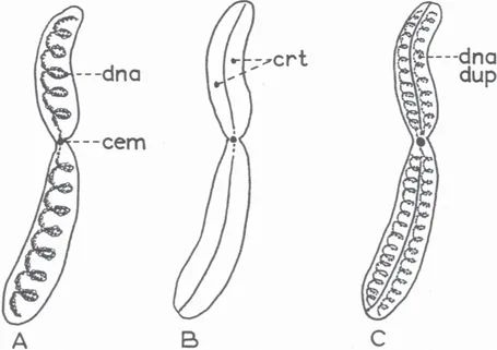
Each chromosome has a constriction somewhere along its length. This is called the centromere and is constant in position for any particular chromosome (Fig. 1.3). The centromere plays a crucial part in cell division by orientating the chromosomes in the correct position in the cytoplasm. When a cell receives a signal to divide, a series of events is set in motion which begin in interphase but soon pass into the stage of prophase.
Figure 1.3: The structure of a chromosome and chromatid. A: Chromosome showing DNA content. B: Chromosome splitting to form chromatids. C: Chromatids containing duplicated DNA. cem = centromere; crt = chromatids; dna = deoxyribosenucleic acid.
Prophase (Initial or First Phase)
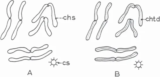
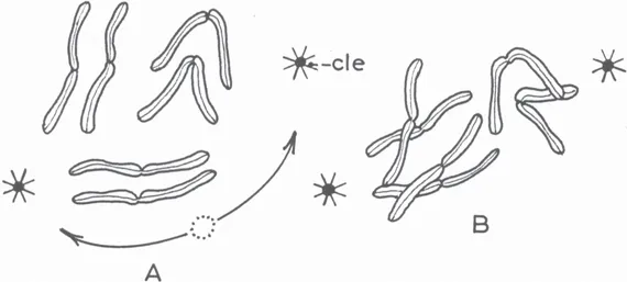
The chromatic material (coloured material of the nucleus) becomes condensed so that the chromosomes are clearly visible (under the microscope) (Fig. 1.4). The chromosomes are set in homologous pairs and soon each chromosome separates longitudinally into a pair of parallel chromatids. At this time the DNA undergoes duplication. The chromatids gradually separate from one another but remain attached at the centromere. At the same time, the centrosome in the cytoplasm divides into two centrioles which move to the opposite poles of the cytoplasm (Fig. 1.5A). When they reach these positions, cytoplasmic ‘rays’ form around them. These are called asters (stars). The nuclear membrane now disintegrates and disappears leaving the chromosomes as a tangled mass in the cytoplasm, between the centrioles (Fig. 1.5B). The rays of the asters around the centrioles extend across the cytoplasm passing between the chromosomes, to form a spindle (Fig. 1.6A). When this process is completed, the cell division enters the next descriptive stage – metaphase.
Figure 1.4: Appearance of condensed chromatin into chromosomes. chs = chromosomes; chtd = chromatids; cs = centrosome.
Figue 1.5: Diagram to illustrate division of centrosome into centrioles as well as formation of asters around centrioles. cle = centriole.
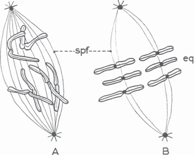
Figure 1.6: Diagram to illustrate formation of the mitotic spindle (A) and attachment of the centromeres to the spindles (B). spf = spindle fibres; eq = equator.
Metaphase (Second Phase)
The chromosomes become shorter and thicker and become arranged on the equator of the spindle. When this occurs, the rays of the spindle become attached to the centromeres of the chromosomes (Fig. 1.6B). It is clear from the configuration of the cell content that the rays of the spindle are ready to pull the mixed chromatids to the opposite ends of the cell. With completion of these events, the division passes to the next stage – anaphase.
Anaphase (Third Phase)
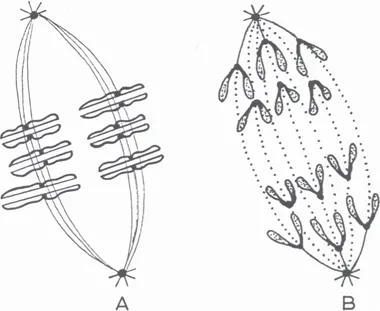
The centromere of each chromosome now splits and the chromosomes also split lengthwise. Being attached to the strands of the spindle, the ‘daughter’ chromosomes (chromatids) are separated from one another and are drawn towards the centrioles (Fig. 1.7A,B). As the chromosomes move towards the poles of the cell, the cell membrane develops a circumferential groove, the cleavage furrow, and at the same time the central part of the spindle becomes narrowed (Fig. 1.8A). With the completion of these events, the division process enters its final stage – telophase.
Figure 1.7: A: Division of chromosome into two chromatids. B: movement of the chromatids to the poles of the spindle.
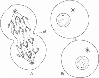
Figure 1.8: Diagram to illustrate the process of division of one cell into two cells. cf = cleavage furrow.
Telophase (Terminal Phase)
The chromosomes lose their crisp and orderly shape and revert to a disorderly mass of chromatin (similar to that of interphase). A nuclear membrane regenerates around each newly formed chromatin mass as the cleavage furrow deepens until the resulting two daughter cells are completely separated from one another. A small concentrated mass of chromatin appears among the chromosomes as the nucleolus and the centrioles revert to a centrosome (Fig. 1.8B).
Each of these cells will have 2n chromosomes as a result of the original chromosomes duplicating into identical chromatids and these will form the chromosomes of the new cells.
Meiosis
This type of cell division is confined to the sex cells and is characterised by the fact that the number of chromosomes in the ‘daughter-cells’ is reduced to half of the original number. Since the original number of chromosomes is 46 (2n), which is the diploid number, the ‘daughter-cells’ after division will contain the haploid number (1n) or 23 chromosomes.
This chromosomal reduction is necessary to form the gametes (sex cells respons...