![]()
CHAPTER ONE
Physiology of vision
Ramesh Seewoodhary
The aim of this chapter is to explore the physiology of vision. It is divided into six parts to give the reader an understanding of the concept of sight:
Embryological development of the eye
Development of visual perception and mechanisms involved in vision
Mechanism of vision in dim light and bright light
Light detection and dark adaptation
Control of eye movements and binocular vision.
Embryological development of the eye
The development of the eye takes place between week 3 and week 10 and involves ectoderm, neural crest cells and mesenchyme. The neural tube ectoderm gives rise to the retina, the iris and ciliary body epithelia, the optic nerve, the smooth muscles of the iris, and some of the vitreous humour. Surface ectoderm gives rise to the lens, the conjunctival and corneal epithelia, the eyelids, and the lacrimal apparatus. The remaining ocular structures form from mesenchyme.
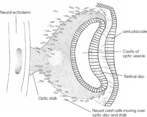
Early eye development starts around day 22 of the embryo’s life, and the eye measures 2–3mm in length. The neural folds fuse to form the neural tube, but, before they complete their closure, the optic sulci appear as shallow grooves or pits in the inner part of the neural folds (Figure 1.1a).
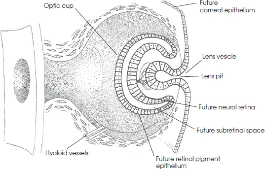
The folds fuse shortly afterwards to form the forebrain, and the optic sulci evaginate to form the optic vesicles (Figure 1.1b). Invagination of the lower surface of the optic stalk and the optic vesicle occur simultaneously, creating a groove known as the choroidal fissure. At the same time, the lens plate also invaginates to form the lens vesicle. By about 4 weeks, the lens vesicle separates completely from the surface ectoderm to lie free in the rim of the optic cup.
The choroidal fissure allows entrance into the optic stalk of the vascular mesoderm, which eventually forms the hyaloid system. As invagination is completed, the choroidal fissure narrows until it is closed by 6 weeks, leaving one small permanent opening at the anterior end of the optic stalk. Through this opening pass the hyaloid artery by the fourth month and the central retinal artery and vein thereafter (Figure 1.1c).
Figure 1.1a The eye at different gestational ages: at 4 weeks
Figure 1.1b The eye at different gestational ages: at 5 weeks
Figure 1.1c The eye at different gestational ages: at 7 weeks
Figure 1.1d The eye at different gestational ages: at 8 weeks
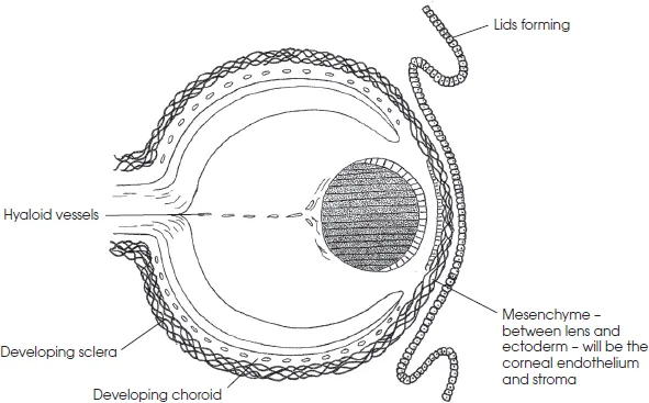
After the fourth month, the general structure of the eye has been determined. Development after this consists of differentiation into individual structures, which occurs more rapidly in the posterior than in the anterior segment early in gestation, and more rapidly in the anterior segment later in gestation (Riordan-Eva & Cunningham 2011).
Generally, the eye is derived from three embryonic layers, namely, surface ectoderm, neural ectoderm and mesoderm
The surface ectoderm gives rise to the lens, the lacrimal gland, corneal epithelium, conjunctiva, adnexal glands and epidermis of the lid.
The neural ectoderm gives rise to the optic vesicle and optic cup, and is responsible for the formation of the retina, ciliary body, pupillary muscle sphincter and dilator, optic nerve and glia.
The mesoderm contributes to the extraocular muscles, sclera, and orbital and ocular vascular endothelium.
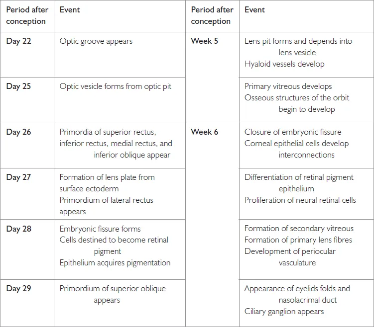
The timeline of ocular embryogenesis is summarised in Table 1.1.
Table 1.1 Timeline of ocular embryogenesis
Healthy eyes and good vision play a critical role in how infants and children learn to see. Poor vision in infants can cause developmental delays. It is important to detect any sight problems early to ensure that babies have the opportunity to develop the visual abilities they need to grow and learn. The next section discusses development of visual perception.
Development of visual perception
Vision, and how the brain uses visual information, are learned skills. Perception of the environment is the end product of the reinterpretation or processing by the visual cortex of the retina’s responses to visual stimuli. There is no strict separation of cortical and retinal function because some processing takes place in the retina and the cortex may not be solely responsible for other processes.
There must be discrimination by means of brightness gradients in the cortex, as well as in the retina. The cortex must be precisely aware of the form and shape of objects and, finally, of the identity of these objects. In the adult, all these processes occur rapidly; we perceive only the objects’ identities and react to them automatically and appropriately, often without being fully conscious of them. In the infant, these processes occur slowly and the final stages of form perception and object identification may not occur at all. Although form perception develops automatically with maturation, identification depends on the capacity to acquire and store information and also on the accumulation of experiences on which identification is based.
Retinal response to light, and to moving objects by reflex responses of blinking, pupillary contraction and eye movements, occurs even in the short-gestation infant and is well established in the normal neonate. According to Gesell et al. (1949), during the first few weeks the infant begins to gaze around and sometimes to fixate objects. Sporadic body activity then ceases and respiration rate alters; there seems to be some awareness of and attention to the environment. This visual exploration of the environment increases with age, especially when infants are handled or situated in complex and unrestricted environments (White & Castle 1964). In young infants the information is fragmentary. Infants aged 2 months cannot store and retain information for more than a very brief period of about 1 second. Each successive appearance of a stimulus is perceived as a new event, so that the environment is experienced in a disconnected manner. At 6 months, the span of attention is so narrow that a single stimulus may capture it entirely. The infant will concentrate on a stimulus in an indiscriminate manner, in order to obtain all possible information from it, and cannot attend selectively. Babies need to learn how to use the visual information the eyes send to the brain in order to understand the world around them and interact with it appropriately (Bowling 2015).
Awareness of an object involves discrimination between it and its surroundings; brightness and colour discrimination also contribute to this. Infants are known to gaze longer at blue and green stimuli and least at yellow. Experiments by Fantz (1958) suggest that, from a very early age, there is some discrimination between patterns. He claimed that within the first week or so of life infants gazed longer at a picture of a face than at a half-black, half-pink shape. It has been found that infants under 1 month tend to look longer at very simple patterns (such as a half-white, half-black field or one with four quadrants) than at more complex patterns. This is a result of the retinal images being too blurred for finer perception.
Accommodation develops during the second month and it is comparable to that of a normal adult by 4 months. Infants look relatively longer at chequered patterns with four squares by week 3. This increases to 64 squares at week 8, and 576 squares at week 14. Attention to pictures and photographs of faces emerges at 3–4 months. Kagan et al. (1966) found that this is followed by the infant smiling more and more, suggesting that some degree of familiarity is involved.
Development in the accuracy of shape perception takes place in close conjunction with tactile exploration. Before the age of 3 months, infants are unaware of any association between the visual and the tactile sensory patterns and do not attempt to handle objects that they perceive visually, or to examine visually the objects that they handle. At 3 months, White and Castle (1964) found that infants stretch out a hand towards an object that they see, and glance rep...