Comprehensively describes bone augmentation techniques and their application to the different anatomical regions of the upper and lower jaws.
Bone Augmentation by Anatomical Region is a unique, evidence-based guide focusing on each specific anatomical region – anterior maxilla, posterior maxilla, anterior mandible, and posterior mandible – in order to emphasize the correct implemented procedures needed to successfully perform oral osseous reconstruction.
Numerous ridge augmentation techniques are covered, including: horizontal and vertical guided bone regeneration, autologous block transplantation, interpositional bone grafting, allogeneic blocks, sandwich technique, split-expansion ridge technique, and sinus floor grafting. Non-augmented approaches such as forced socket site extrusion and the installation of digitally printed implants are also presented and discussed.
Guides readers on tackling bone augmentation via anatomical region of the jaws and their related surrounding muscles, vascularization and innervation
Presents innovative augmentation techniques for the anterior maxilla, posterior maxilla, anterior mandible, and posterior mandible
Includes clinical photographs in each section and a decision tree to help readers select the appropriate surgical modality
Bone Augmentation by Anatomical Region is a specialist resource suitable for dentists who practice implant dentistry, oral surgeons, oral and maxillofacial surgeons, periodontists, and postgraduate dental students in the above-mentioned disciplines. Please note Due to recently developments, part of Chapter 2Biologic Conditions for Bone Growth and Maintenance: Managing the Oxidative Stress has been amended which will be available in all future reprints. All electronic versions have been updated.
Foire aux questions
Comment puis-je résilier mon abonnement ?
Il vous suffit de vous rendre dans la section compte dans paramètres et de cliquer sur « Résilier l’abonnement ». C’est aussi simple que cela ! Une fois que vous aurez résilié votre abonnement, il restera actif pour le reste de la période pour laquelle vous avez payé. Découvrez-en plus ici.
Puis-je / comment puis-je télécharger des livres ?
Pour le moment, tous nos livres en format ePub adaptés aux mobiles peuvent être téléchargés via l’application. La plupart de nos PDF sont également disponibles en téléchargement et les autres seront téléchargeables très prochainement. Découvrez-en plus ici.
Quelle est la différence entre les formules tarifaires ?
Les deux abonnements vous donnent un accès complet à la bibliothèque et à toutes les fonctionnalités de Perlego. Les seules différences sont les tarifs ainsi que la période d’abonnement : avec l’abonnement annuel, vous économiserez environ 30 % par rapport à 12 mois d’abonnement mensuel.
Qu’est-ce que Perlego ?
Nous sommes un service d’abonnement à des ouvrages universitaires en ligne, où vous pouvez accéder à toute une bibliothèque pour un prix inférieur à celui d’un seul livre par mois. Avec plus d’un million de livres sur plus de 1 000 sujets, nous avons ce qu’il vous faut ! Découvrez-en plus ici.
Prenez-vous en charge la synthèse vocale ?
Recherchez le symbole Écouter sur votre prochain livre pour voir si vous pouvez l’écouter. L’outil Écouter lit le texte à haute voix pour vous, en surlignant le passage qui est en cours de lecture. Vous pouvez le mettre sur pause, l’accélérer ou le ralentir. Découvrez-en plus ici.
Est-ce que Bone Augmentation by Anatomical Region est un PDF/ePUB en ligne ?
Oui, vous pouvez accéder à Bone Augmentation by Anatomical Region par Zvi Artzi, Zvi Artzi en format PDF et/ou ePUB ainsi qu’à d’autres livres populaires dans Medicine et Dentistry. Nous disposons de plus d’un million d’ouvrages à découvrir dans notre catalogue.
1 The Anatomy of the Maxilla and the Mandible: Related Structures and Inserted Muscles
Dmitri Lev and Zvi Artzi
The success of oral rehabilitation and related surgical interventional procedures in oral implantology depends upon in‐depth knowledge and understanding of the head and neck anatomy. Anatomical structures in the head and neck region are numerous and densely packed in a relatively small volume. These organs are designed to serve various systems, such as masticatory, olfactory, lacrimal, visual, and others. Overlapping of these systems, however, makes it almost impossible to draw clear demarcation lines between them.
This chapter will focus mainly on the oral anatomy, and additionally consider the structures which are topographically and functionally part of the oral apparatus.
Bones
The mandible, maxilla, and palatine bones form the boundaries of the oral cavity. Because the number of teeth changes during an individual's lifetime, the mandible and maxilla, more than the other bones of the viscerocranium, are permanently and extensively developed and modified during childhood and adolescence, only to undergo significant degeneration with aging.
Maxilla
Each maxilla is composed of two bones, the maxilla proper and the premaxilla, which fuse during the last trimester of fetal development (Figure 1.1). The incisive suture connecting the maxilla proper and premaxilla is located on the inferior surface of the hard palate, and it becomes obliterated in varying degrees during midlife. Two maxillae form the entire upper jaw and most of the middle face. Maxillary occupation of the central part of the facial skeleton involves the oral, nasal, and orbital cavities, and their articulation with the ethmoid, frontal, lacrimal, nasal, inferior nasal concha, vomer, palatine, and opposite maxilla bones. Its hollowed pyramidal‐shaped body includes four processes (see Table 1.1) which radiate from the maxilla in directions corresponding to the buttress lines of the viscerocranium.
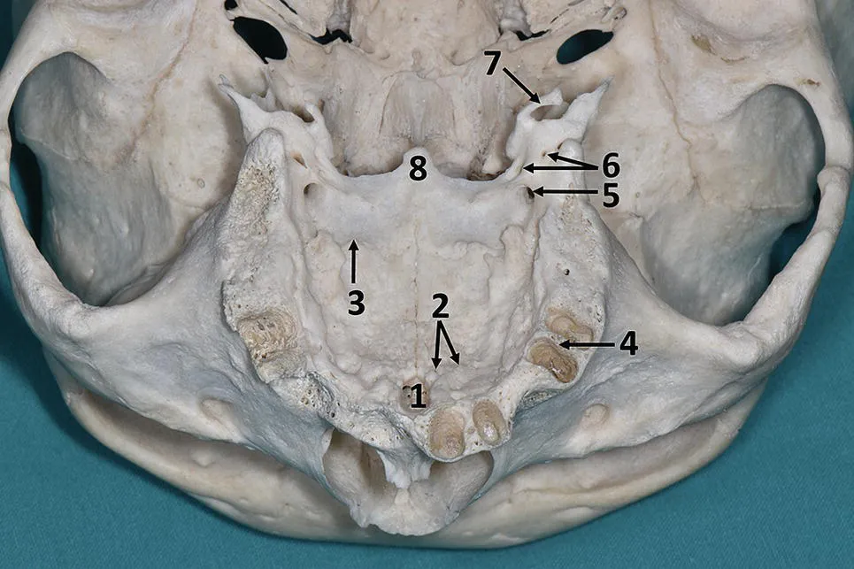
The long frontal process ascends between the lacrimal and nasal bones to articulate with the frontal bone via the frontomaxillary suture. The short zygomatic process protrudes laterally and connects with the maxillary process of the zygomatic bone via the zygomaticomaxillary suture. The ridge on the inferior aspect of the zygomatic process separates the anterior and posterior concavities: the former continues up to the anterolateral surface of the maxillary body, while the latter terminates opposite the infratemporal fossa. The alveolar process (Figure 1.1) descends from the anterolateral and posterior maxilla body surface. It supports the teeth and gradually becomes wider in a posterior direction. The alveolar process is composed of one external and one internal cortical plate, and a considerable amount of trabecular bone tissue is sandwiched between them. Posteriorly, the cortical plates are united. The inferior edge of the alveolar process is deeply grooved, and the cortical plates are interconnected by perpendicular interalveolar septa, which divide the groove into eight alveolar sockets in the adult maxilla. In the posterior/distal three sockets, the inter‐radical septa separate between the individual roots in multirooted teeth. The inter‐radical septum of the first premolar socket is parallel to the alveolar/cortical plates. The configuration and size of the roots determine the alveolar socket morphology. Tooth extraction causes gradual resorption of the alveolus. The palatine process originates from the border between the anterior two‐thirds of the maxilla and its alveolar process. It projects medially to meet its fellow palatine process at the intermaxillary suture. The posterior edge of the palatine process connects with the horizontal process of the palatine bone at the transverse palatine suture. The horizontal plate of the palatine process forms a right angle with the posterior aspect of the alveolar process. The angle is poorly defined anteriorly, and the oral surface of the palatine process slopes down at this point. The concave, rough undersurface of the palatine process provides firm attachment of the masticatory mucosa. The superior nasal surface is also concave but smooth, and the mucosa is loosely attached to it. Two palatine processes have an elevation along the intermaxillary suture, which forms the nasal crest for attachment of the vomer bone. The prominent anterior part of the nasal crest (the “incisor spine”) is the site of attachment of the cartilaginous nasal septum. The nasopalatine canal traverses the palate just posteriorly to the incisor spine.
The nasopalatine canal is usually described as a Y‐shaped channel starting from two Stenson's foramina on the nasal surface of both palatine processes and ending inferiorly as a single opening on the oral roof on the bottom of the incisive fossa, just posterior to the central incisors. However dominant, this arrangement is present in only fewer than 50% of the population. The nasopalatine canal may contain from one to four channels between the superior and inferior openings (Song et al. 2009). Within the nasopalatine canal, the nasopalatine nerve communicates with the greater palatine nerve, and the greater (descending) palatine artery anastomoses with the posterior septal branch of the sphenopalatine artery.
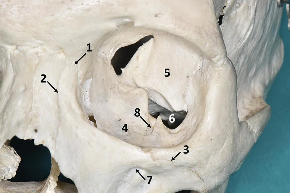
Figure 1.2 Orbit (anterior view). 1 – Frontomaxillary suture, 2 – nasomaxillary suture, 3 – zygomaticomaxillary suture, 4 – superior surface of the maxillary body, 5 – greater wing of the sphenoid bone, 6 – infraorbital fissure, 7 – infraorbital foramen, 8 – infraorbital groove.
The hollowed maxillary body has a pyramidal shape. The pyramid a...