eBook - ePub
Legionellosis
Volume II
Katz
This is a test
- 221 pages
- English
- ePUB (adapté aux mobiles)
- Disponible sur iOS et Android
eBook - ePub
Legionellosis
Volume II
Katz
Détails du livre
Aperçu du livre
Table des matières
Citations
À propos de ce livre
Legionellosis is a text in two volumes that presents the modern viewpoint of the agent and the disease. It also chronicles the history of the discovery of Legionella pneumophila. Volume 1 discusses current aspects of the microbe including taxonomy, morphology, biochemistry, and physiology. It also discusses the illness including clinical features, pathology, and therapy. Volume II details the laboratory diagnosis, epidemiology, and pathology. The contributors are amongst the most eminent scientists in their respective fields.
Foire aux questions
Comment puis-je résilier mon abonnement ?
Il vous suffit de vous rendre dans la section compte dans paramètres et de cliquer sur « Résilier l’abonnement ». C’est aussi simple que cela ! Une fois que vous aurez résilié votre abonnement, il restera actif pour le reste de la période pour laquelle vous avez payé. Découvrez-en plus ici.
Puis-je / comment puis-je télécharger des livres ?
Pour le moment, tous nos livres en format ePub adaptés aux mobiles peuvent être téléchargés via l’application. La plupart de nos PDF sont également disponibles en téléchargement et les autres seront téléchargeables très prochainement. Découvrez-en plus ici.
Quelle est la différence entre les formules tarifaires ?
Les deux abonnements vous donnent un accès complet à la bibliothèque et à toutes les fonctionnalités de Perlego. Les seules différences sont les tarifs ainsi que la période d’abonnement : avec l’abonnement annuel, vous économiserez environ 30 % par rapport à 12 mois d’abonnement mensuel.
Qu’est-ce que Perlego ?
Nous sommes un service d’abonnement à des ouvrages universitaires en ligne, où vous pouvez accéder à toute une bibliothèque pour un prix inférieur à celui d’un seul livre par mois. Avec plus d’un million de livres sur plus de 1 000 sujets, nous avons ce qu’il vous faut ! Découvrez-en plus ici.
Prenez-vous en charge la synthèse vocale ?
Recherchez le symbole Écouter sur votre prochain livre pour voir si vous pouvez l’écouter. L’outil Écouter lit le texte à haute voix pour vous, en surlignant le passage qui est en cours de lecture. Vous pouvez le mettre sur pause, l’accélérer ou le ralentir. Découvrez-en plus ici.
Est-ce que Legionellosis est un PDF/ePUB en ligne ?
Oui, vous pouvez accéder à Legionellosis par Katz en format PDF et/ou ePUB ainsi qu’à d’autres livres populaires dans Ciencias biológicas et Microbiología. Nous disposons de plus d’un million d’ouvrages à découvrir dans notre catalogue.
Informations
Part I: Laboratory Diagnosis of Legionellosis
Chapter 1
Diagnosis: Culture
James C. Feeley
Table Of Contents
I. Introduction
II. Development of Media
III. Recommended Medium
A. BCYE α Medium
1. Composition
2. Sources for Ingredients
3. Directions for Preparation
4. Precautions
5. Quality Control
6. Antibiotic Supplementation
B. BCYE α Diphasic Blood Culture Medium
1. Composition
2. Preparation
IV. Specimens
A. Safety Considerations
B. Types
C. Collection and Transport
V. Culture And Primary Isolation
VI. Examination and Identification of Cultures
References
I. Introduction
The diagnosis of acute cases of Legionnaires’ disease has been primarily made clinically. Laboratory confirmation followed days or weeks later. However, this is changing with the development of rapid antigen detection tests9 and the improvement of the culture media by addition of ACES buffer,” a-ketoglutarate,4 and effective antimicrobials as selective agents1,24,13 to the charcoal-yeast extract (CYE) base medium.7 In the U.S., the sensitivity of culture currently ranges between 50 to 80% in laboratories that routinely culture specimens for Legionella. Microbiologists in these laboratories can detect Legionella as early as 2 days of incubation following inoculation of the media by picking microcolonies using a dissecting microscope, and staining them with immunofluorescent antibody reagents.
II. Development of Media
Since an understanding of the growth requirements of Legionella species has proven helpful to many microbiologists in isolating Legionella species, a brief history of the development of the media is provided. The Legionnaires’ disease bacterium (LDB), now called L. pneumophila, was first isolated by using rickettsial techniques.10 Guinea pigs were inoculated intraperitoneally with lung tissue from fatal cases of Legionnaire's disease. When a Guinea pig became ill, its spleen was removed, ground, and suspended in sterile phosphate buffered saline and inoculated into yolk sacs of embryonated hens eggs. The small Gram-negative rods in the yolk sacs that were identified as the etiologic agent of Legionnaires’ disease by indirect immunofluorescent staining grew only on Mueller-Hinton supplemented with 1 % hemoglobin and 1 % IsoVitalex® (MH-IH) agar but did not grow on 16 other types of media.5 Feeley et al. examined components of the MH-IH medium5 to determine what special requirements that LDB needed for growth.
They found that MH-IH agars made without l-cysteine HCL and iron salts were unable to support the growth of LDB, that pH 6.9 was optimal for “growth initiation” for LDB on primary isolation, and that cultures maintained on bacteriological media were less affected by pH. These observations have been applied in the development of most of the media designed specifically to grow L. pneumophila and in the development of quality control testing procedures for Legionella.
Feeley-Gorman (F-G) agar was the first medium that was specifically formulated to grow L. pneumophila. It was substantially more sensitive than MH-IH agar, and it also enabled detection of the characteristic browning that most Legionella species produce on tyrosine containing media. Unfortunately, the Mueller-Hinton basal medium used in making F-G agar varied widely from lot to lot in its ability to support growth of L. pneumophila. For this reason and the fact that F-G agar does not support primary isolation of non-pneumophila Legionella species, it should not be used for primary isolation. CYE agar was developed to replace F-G agar.7 It was found to be 100 times more sensitive, to support faster growth, and to support primary isolation of all Legionella species. However, one shortcoming that CYE agar was found to have was its inability to recover Legionella from specimens such as sputa that contain other flora. One possible explanation for this was that the other flora produce substances such as bacteriocins that were inhibitory to Legionella. Another explanation was that the other flora rapidly changed the pH of the agar from 6.9 before Legionella could initiate growth. The addition of ACES buffer” was added to correct this fault by increasing the buffering capacity of CYE agar. The new medium that resulted was called “buffered charcoal yeast extract” (BCYE) agar and did support better growth of Legionella from all kinds of specimens. Another improvement of the basal medium resulted with the addition of α-ketoglutarate4 to BCYE agar. This enabled faster initiation of growth of Legionella as well as giving higher recovery of Legionella from antibiotic containing media supplemented with it as contrasted to lower recovery of Legionella from antibiotic containing media not supplemented with it. BCYE media containing α-ketoglutarate is designated BCYEα agar.
III. Recommended Medium
BCYEα agar and BCYEα diphasic blood culture medium are the best basal media currently available for culturing Legionella. BCYEα agar can be made selective by supplementation with various antimicrobials.1,24,13 Some investigators have added dyes12,13 to enable detection and differentiation of Legionella species. This has proved extremely helpful in examining water from hospitals contaminated with two or more species of Legionella such as L. pneumophila and L. miedadei. However, dye incorporation is not recommended for general usage because it complicates the medium and may inhibit some species of Legionella. Many investigators prefer using DFA staining for detecting the numerous species that may be in a specimen. Many commercial media companies are now selling BCYEα agar with and without antimicrobial and dye supplementation.
A. BCYEα Medium
1. Composition
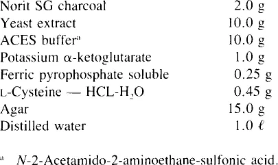
2. Sources for Ingredients
All of the ingredients of BCYEα agar are now commercially available and most are easily obtained, including Norit SG charcoal and ferric pyrophosphate soluble. Sigma Chemical Co., St. Louis, Mo., sells both, which are listed, respectively, as products C5510 and P6526. Ferric pyrophosphate soluble can now also be purchased from City Chemical Corp.. 132 West 22nd St., New York, N.Y. 10011, and is listed as product number F454. Although some agars have been found toxic to Legionella; agars from DIFCO and GIBCO and OXOID have been found to be suitable for use and should be added in amounts (grams per liter) suggested by the individual manufacturer.
3. Directions for Preparation
The yeast extract and some of the other components of BCYEα agar can be easily denatured during preparation of the medium. The following directions are provided to prevent this:
- Add 940 ml of distilled water to 10 g ACES buffer and dissolve by warming in a 50°C water bath. Since the buffer does not come to its pKa of 6.9 and is very acid, it needs to be adjusted to pH 6.9 by adding approximately 40 to 43 ml of 1 N KOH to the buffer solution. Note well that NaOH must not be used to adjust the ACES buffer because it is inhibitory to L. pneumophila in the final medium.
- Pour the buffer solution into a second flask that contains the dry powders of charcoal, yeast-extract, agar-agar, and α-ketoglutarate, and mix well.
- Gently heat the mixture to dissolve the agar-agar and sterilize it by autoclaving at 15 lb for 15 min. The medium must not be subjected to prolonged heating because it is extremely sensitive to high temperatures.
- Cool the medium immediately to 50°C by placing the flask in a 50°C water bath.
- For medium intended to be complete (that containing l-cysteine), add 10 ml of a filter sterilized solution of lcysteine (0.45 g/10 ml) and mix thoroughly. l-Cysteine should not be added to media that will be used to test l-cysteine requirement of cultures. This medium should be labeled “C-” for “Cysteine minus” BCYE agar.
- Add separately 10 ml of a filter-sterilized solution of soluble ferric pyrophosphate (0.25 g/10 ml), and mix thoroughly. The iron and cysteine solutions should not be mixed prior to addition to medium because a toxic complex may result.
- The final pH of the medium should be 6.90 ± 0.05 at 20°C. The pH of the final medium can be adjusted while the medium is being held at 50°C in the water bath. At this temperature the medium should be adjusted to 6.30. The reason is that the pKa of ACES buffer changes — 0.02 pH units per + 1°C greater than 20°C. On cooling to room tem...