
eBook - ePub
Introduction to Practical Ore Microscopy
P.R. Ineson
This is a test
Partager le livre
- 192 pages
- English
- ePUB (adapté aux mobiles)
- Disponible sur iOS et Android
eBook - ePub
Introduction to Practical Ore Microscopy
P.R. Ineson
Détails du livre
Aperçu du livre
Table des matières
Citations
À propos de ce livre
First published in 1989. This book is designed to be an introduction to Ore microscopy, which is the traditional name for the study of opaque minerals using a polarising 'incident light' microscope. It is also known as reflected light microscopy. It has applications in the fields of mineralogy, economic geology, mineral dressing, metallurgy and in the study of igneous, metamorphic and sedimentary rocks which contain opaque minerals. Although mineral identification is an important aspect of the study of the opaque ores, an examination of the textures and structures is also valuable.
Foire aux questions
Comment puis-je résilier mon abonnement ?
Il vous suffit de vous rendre dans la section compte dans paramètres et de cliquer sur « Résilier l’abonnement ». C’est aussi simple que cela ! Une fois que vous aurez résilié votre abonnement, il restera actif pour le reste de la période pour laquelle vous avez payé. Découvrez-en plus ici.
Puis-je / comment puis-je télécharger des livres ?
Pour le moment, tous nos livres en format ePub adaptés aux mobiles peuvent être téléchargés via l’application. La plupart de nos PDF sont également disponibles en téléchargement et les autres seront téléchargeables très prochainement. Découvrez-en plus ici.
Quelle est la différence entre les formules tarifaires ?
Les deux abonnements vous donnent un accès complet à la bibliothèque et à toutes les fonctionnalités de Perlego. Les seules différences sont les tarifs ainsi que la période d’abonnement : avec l’abonnement annuel, vous économiserez environ 30 % par rapport à 12 mois d’abonnement mensuel.
Qu’est-ce que Perlego ?
Nous sommes un service d’abonnement à des ouvrages universitaires en ligne, où vous pouvez accéder à toute une bibliothèque pour un prix inférieur à celui d’un seul livre par mois. Avec plus d’un million de livres sur plus de 1 000 sujets, nous avons ce qu’il vous faut ! Découvrez-en plus ici.
Prenez-vous en charge la synthèse vocale ?
Recherchez le symbole Écouter sur votre prochain livre pour voir si vous pouvez l’écouter. L’outil Écouter lit le texte à haute voix pour vous, en surlignant le passage qui est en cours de lecture. Vous pouvez le mettre sur pause, l’accélérer ou le ralentir. Découvrez-en plus ici.
Est-ce que Introduction to Practical Ore Microscopy est un PDF/ePUB en ligne ?
Oui, vous pouvez accéder à Introduction to Practical Ore Microscopy par P.R. Ineson en format PDF et/ou ePUB ainsi qu’à d’autres livres populaires dans Sciences physiques et Géographie. Nous disposons de plus d’un million d’ouvrages à découvrir dans notre catalogue.
Informations
1 INTRODUCTION
Ore microscopy is the traditional name for the study of opaque minerals using a polarising ‘incident light’ microscope. It is also known as reflected light microscopy. It has applications in the fields of mineralogy, economic geology, mineral dressing, metallurgy and in the study of igneous, metamorphic and sedimentary rocks which contain opaque minerals. Although mineral identification is an important aspect of the study of the opaque ores, an examination of the textures and structures is also valuable.
This book is designed to form an introduction to the subject. The reader who wishes to study the subject in greater depth is referred to the more comprehensive texts by Cameron (1961), Shouten (1962), Freund (1966), Galopin and Henry (1972), Uytenbogaardt and Burke (1973), Ramdohr (1980), Craig and Vaughan (1981) and Picot and Johan (1982). However, the principal methods of identification and the most commonly noted textures are here described for a selection of minerals which should suffice for most students. Company geologists can examine either run-of-the-mill or exotic mineral grades, whereas students at universities, technical colleges, mining schools, museums and similar institutions normally see only exotic or high-grade samples, and although these can be helpful in ‘learning the trade’ they can be a misleading preparation for operational conditions. This has been borne in mind in making the selection of minerals to be dealt with in the present text.
Ore microscopy and the use of the reflected light microscope with its varied ancillary equipment cannot be considered to be a comprehensive study. It frequently needs to be combined with other investigations, e.g. chemical analyses (by spectrographic, X-ray fluorescence, electron microprobe, atomic absorption, wet chemical and other techniques), X-ray diffraction, universal stage, and grain size measurements. In general, ore microscopic examination is a useful, or even an essential, precursor to such investigations. Likewise, the ore minerals cannot be fully comprehended without a knowledge of the deposits in which they occur. There are many suitable texts which cover this aspect, but the following may be found to be appropriate: Park and MacDiarmid (1964), Brown (1967), Stanton (1972), Baumann (1976), Wolf (1976), Barnes (1979), Evans (1980), Jensen and Bateman (1981) and Hutchison (1983).
Mineralogical texts are also plentiful but the following may prove to be sufficient at this initial level of study: Deer, Howie and Zussman (1962), Ribbe (1974), Rumble (1976) and Vaughan and Craig (1978).
2 THE ORE MICROSCOPE
2.1 GENERAL COMMENTS
The microscope (Fig. 2.1) used for the study of ‘ore’ or ‘opaque’ minerals is basically the same as a petrological microscope except that it uses reflected light. (It should not be assumed, however, that experience with the petrological microscope can be directly transferred to the use of the reflected-light microscope.) Some modern microscopes (Fig. 2.2) may be dual purpose and have a combination of both transmitted and reflected light sources built into their assembly. These are capable of examining polished samples, petrological thin sections and polished thin sections. They are particularly useful when both the opaque and nonopaque minerals have to be examined for their mutual relationships and textures, e.g. ore minerals together with gangue or the host/wallrock minerals.
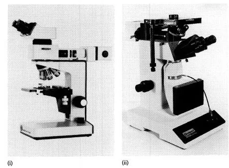
Fig. 2.1 Reflected/incident light (ore) microscopes. (i) Leitz ‘Laborlux 12ME’. Reproduced by courtesy of Ernst Leitz Wetzlar GmbH. (ii) Union ‘Unimet’ inverted stage microscope with binocular head. Reproduced by courtesy of Vickers Instruments Ltd.
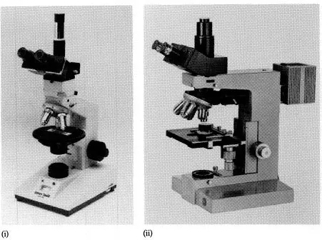
Fig. 2.2 Dual-purpose microscopes capable of viewing samples in transmitted as well as reflected/incident light. (i) ‘MP3500’ James Swift Polarising Microscope with trinocular head. Reproduced by courtesy of Prior Scientific Instruments Ltd. (ii) Leitz ‘Metallux II’ microscope. Reproduced by courtesy of Ernst Leitz Wetzlar GmbH.
The microscope should possess:
1. A rotatable stage.
2. One or more objective lenses of either the ‘dry’ (or air) type or the ‘oil’ immersion type.
3. An ocular (eyepiece) lens.
4. Polariser and analyser.
5. A reflector.
6. A light source, preferably built-in and stabilised.
It may also have the following attachments or accessories:
1. A sample holder (various models are available).
2. A mechanical stage.
3. An eyepiece micrometer.
4. A photometer.
5. A monochromator and/or filters.
6. A camera attachment.
2.2 THE ROTATABLE STAGE
The stage (Fig. 2.3) must be perpendicular to the light path and centred relative to the objectives. Once correctly aligned, it does not normally need any further adjustments. Angular measurements are possible with the verniers often provided. A mechanical stage may be fitted for the systematic traversing of a sample in the X and Y directions.
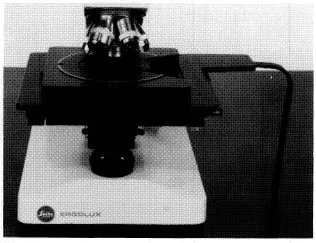
Fig. 2.3 The Leitz ‘Ergolux’ Microscope stage. Reproduced by courtesy of Ernst Leitz Wetzlar GmbH.
2.3 THE OBJECTIVE LENS(ES)
These may be classified as: achromatic, apochromatic and fluorite or semi-apochromatic and are applicable for both air and oil immersion. They may also be made to give a flat primary image and as such are prefixed ‘flat-field’ or ‘plan’ and, if so, are very useful for examining large fields of view and when photographing the sample. The magnification (degree of enlargement) can vary from ×2 to ×125 or greater. The numerical aperture (NA) indicates the ability of the lens to distinguish fine structural detail and determines the depth of field and the useful range of magnification. A typical lens ‘turret’ is shown in Fig. 2.4.
Achromatic lenses are the most common and the cheapest. They are corrected for spherical aberration for one colour, normally yellow-green, and for chromatic aberration for two colours. When used with white light, colour fringes may be seen in the outer image margins. If black-and-white microphotography is used with these lenses, fuzziness is possible. In monochromatic light, the image is sharp.
Apochromatic lenses are better but more expensive. They are corrected for spherical aberration for two colours (blue and green) and for chromatic aberration for the primary spectral colours. They produce a sharper image and should give a better print in colour photomicroscopy. To give an even better performance, they should be used with ‘compensating’ eyepieces.
Fluorite objective lenses are a compromise between achromatic and apochromatic lenses, and should be used with compensating eyepieces.
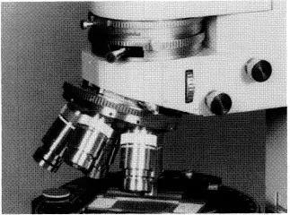
Fig. 2.4 A five-lens microscope turret fitted to a ‘M17’ Vickers Microscope. Reproduced by courtesy of Vickers Instruments Ltd.
Most lenses (Fig. 2.5) of a low to medium magnification are ‘dry’ lenses, and as such are designed to be used when air is the medium between the sample and the objective lens and/or when the specimen is covered with a glass. Though they are frequently used in transmitted light studies, in reflected light microscopy they may give distorted images. Immersion objective lenses, which are manufactured to give all possibly required magnifications, should be the most commonly used type in reflected light studies and especially so when high resolution and magnification are demanded. They are, however, not often used by persons with little experience because they require frequent and careful cleaning of the lens (to stop it gathering dust) each time either the lens or the sample is changed. The immersion medium may be oil or water, but it is normally oil with a refractive index of 1.515. These special lenses reduce the reflectance of the mineral, increase the colour differences, reduce the diffuse light scattering and permit the observation of weak anisotropism and bireflectance.
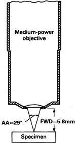
Fig. 2.5 Sketch of a typical lens used in ore microscopy. It shows the free working distance (FWD) and angular aperture (AA). Reproduced from An Introduction to the Methods of Optical Crystallography by F.D. Bloss (1961); Holt, Rinehart and Winston Inc.
2.4 THE OCULAR (EYEPIECE) LENS(ES)
Monocular and binocular eyepieces are available for most microscopes. If possible, to alleviate eye strain, binocular eyepieces should be used and the position of the two eyepieces adjusted so that a single perfectly circular field of view is observed. The oculars are normally of × 5 and × 10 magnification, with or without a perpendicular crosshair, but may be fitted with a micrometer disc or grid for the accurate measurement of parts of the sample. These internally fitted attachments are capable of being brought into focus by rotating the eyepiece. This should always be one of the very first operations to be undertaken. Photomicroscopy eyepieces do not, normally, have crosshairs and are of the compensating type in order to correct for chromatic aberration.
2.4.1 CARE AND CLEANING OF LENSES
The objective and ocular lenses are prone to gathering dust as well as accidentally touching the sample or your eyes. They may thus become ‘dirty’ or produce an effect called ‘blooming’. The lenses should not be cleaned with ordinary tissues or cloths, or breathed upon, since this apparently rapid and easy remedy may cause one of the most expensive pieces of the microscope, i.e. the lens, to be ruined. The recommended lens-cleaning tissues should be used or assistance requested from an experienced person.
2.5 THE POLARISER AND ANALYSER
The polariser is normally located between the light source and the objective lens and often between two sets of diaphragms. It is usually a polaroid plate which, is capable of being easily rotated. It is mounted perpendicular to the analyser. The polarisation effects are often more easily noted if the polars are a few degrees from the true 90° position, and this is especially so for anisotropic minerals if they occur in a groundmass or matrix of a more strongly anisotropic mineral. This effect may be achieved by slightly (3–5°) rotating either the polariser or the analyser from the totally crossed position.
Rather than rotating the stage, for the movement of the whole sample may distract the eyes, it is better or more advisable to rotate the analyser or polariser about the extinction position. The presence or absence of anisotropism in the grain may thus be confirmed. If need be, adjust one of the diaphragms, in order to view only that grain.
2.6 THE REFLECTOR
The reflector, which is either a glass plate or a prism, is the means by which the light is brought vertically onto the polished samples surface. A glass plate reflector only allows about 25% of the illuminating light that first reached the reflector to ultimately reach the ocular lens. A prism reflector has approximately 50% efficiency. If the sample is viewed in a conoscopic field of view, only half of the polarisation figure will be seen, for the other half is occupied by the prism. Normally these reflectors are fixed and adjustment is a specialised operation.
2.7 THE LIGHT SOURCE (ILLUMINATION, ILLUMINATOR)
Possibly one of the most frequent errors in ore microscopy is not to pay sufficient attention to the alignment of the light source.
This aspect cannot be overst...