![]()
1
Emergency echocardiography General considerations
Ivan Stankovic, Andreas Hagendorff, and Aleksandar N. Neskovic
Key points
• Emergency echocardiography is a comprehensive diagnostic procedure performed by cardiologists or adequately trained noncardiologists who are able to independently perform and interpret the study.
• Echocardiography in the emergency setting is a highly demanding procedure, and because of the serious implications of the examination results, it should not be attempted by inexperienced health care providers without supervision.
• Standards are proposed for ultrasound equipment, execution, documentation, and interpretation, as well as for education and training of physicians performing echocardiography in the emergency setting.
• Acute chest pain, acute dyspnea, hemodynamic instability, new murmur, syncope, chest trauma, and cardiac arrest are the main clinical situations in which emergency echocardiography is required.
Rapid and accurate evaluation of unstable patients presenting with symptoms suggestive of cardiovascular pathology is a crucial task in a busy emergency department (ED). A focused history taking, physical examination, and 12-lead electrocardiogram (ECG) remain essential first steps of guideline-proposed algorithms, but additional laboratory tests and imaging studies are required in a sizeable number of patients. Among the currently available imaging techniques, echocardiography seems perfectly fitted for demanding emergency settings because of its availability, portability, and accuracy. It can be performed promptly virtually everywhere, and the results of examination are immediately available, allowing initiation of appropriate treatment without unnecessary delay.
It should be noted that although requirements and recommendations proposed in this chapter are based on international guidelines,1,2 models for use of emergency echocardiography in everyday clinical practice depend on local human and technological resources and may differ among institutions and countries.
In this chapter, we provide a brief overview of practical and medicolegal aspects pertinent to emergency echocardiography1; specific considerations related to pocket-size imaging devices and focused cardiac ultrasound (FoCUS) in the emergency setting, as well as echocardiographic features of particular cardiac emergencies, are detailed in corresponding chapters of this book (Chapters 18 and 19).
Terminology
The current trend of rapidly increasing use of echocardiography in emergency settings by noncardiologists or cardiologists without specific expertise1 occurs in parallel with an evolving trend of using the cardiac ultrasound examination as a bedside, point-of-care diagnostic test in emergency settings. This examination has been named focused cardiac ultrasound. It is important to distinguish emergency echocardiography and FoCUS for practical, logistic, educational, and medicolegal reasons.3
Echocardiography is a comprehensive investigation requiring maximum technical skills along with expertise in cardiovascular pathophysiology and cardiovascular diseases. Thus, the term echocardiography refers to comprehensive standard echocardiography in emergency settings—that is, emergency echocardiography, which always represents a full echocardiographic investigation of cardiac morphology and function, using fully equipped echocardiographic machines, performed by a sufficiently trained operator who is able to independently perform and interpret the study results.1,3
The term FoCUS defines the point-of-care cardiac ultrasound examination, performed according to a standardized but restricted scanning protocol to add information to the physical examination findings, by an operator who is not necessarily fully trained in echocardiography but rather appropriately trained in FoCUS, and who is at the same time usually responsible for immediate decision making and/or treatment.3
Importantly, both cardiologists and noncardiologists can perform either echocardiographic examinations or FoCUS, depending on clinical circumstances, existing equipment, and expertise. Thus, although FoCUS is typically used by noncardiologists who have undergone minimal training, it can also be performed by fully trained cardiologists in emergency settings.
Echocardiography in the emergency setting
Even under the best possible conditions for the evaluation of patients, echocardiography is a highly operator-dependent technique, and human factors (i.e., ability, training, and experience) account for the vast majority of diagnostic errors. In the stressful emergency situation, critically ill patients are typically scanned in minimal time, and potentially catastrophic errors are even more likely to occur, especially if the examination is performed by an inexperienced operator. Echocardiography is already a widely available imaging modality, but with the advent of pocket-size imaging devices, even wider dissemination of the technique is expected, with increasing use by non-experts (emergency physicians, intensivists, anesthesiologists, cardiac surgeons). Medical professionals involved in emergency echocardiography must be able not only to obtain adequate images under challenging conditions but also to interpret them accurately. A failure to either obtain the appropriate image or understand what is imaged may result in misleading and dangerous conclusions. A failure of inexperienced echocardiographers to distinguish normal variants and artifacts from serious pathology may result in unnecessary hospital admissions and further costly investigations (Figure 1.1) (V1.1, V1.2A, and V1.2B), and serious pathology can be missed because of suboptimal scanning technique (Figure 1.2 and V1.3A and V1.3B).
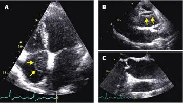
Figure 1.1
Challenging image interpretation in patients with suspected cardiac emergencies. (A) Unusually prominent Chiari’s network (an embryologic remnant normally seen in approximately 2% of the population; arrows) was initially interpreted as right heart thrombus (V1.1). (B) Ultrasound artifact mimicking intimal flap in the ascending aorta on transthoracic examination (arrows, see V1.2) required transesophageal study (C) to rule out aortic dissection (V1.3). (Figures 1.1B and 1.1C provided by VC.)
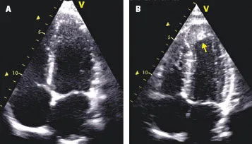
Figure 1.2
Challenging image acquisition in the emergency setting. Important findings may be overlooked because of poor scanning technique resulting in apical foreshortening (A) (see V1.3A). Apical dyskinesia and thrombus (arrow) became obvious only when a full-length image of the left ventricle was obtained (B) (see V1.3B).
Apart from difficulties in image acquisition and interpretation, noncardiologists performing emergency echocardiography may also be challenged by lack of appropriate knowledge that is required to put echocardiographic findings in the clinical context. A pattern recognition approach is useful for diagnosing obvious pathology, such as pericardial effusion or severe left ventricular (LV) dysfunction, whereas the assessment of complicated emergency cases (e.g., severe hypotension caused by dynamic LV tract obstruction) requires the comprehensive understanding of cardiovascular pathology to establish the underlying cause of the patient’s instability.
Table 1.1 “ABCD approach” in performing emergency echocardiography
A | Awareness | • Fight against routine • Think beyond apparent explanations |
B | Be Suspicious | • Referral diagnosis may be misleading • Never trust, confirm |
C | Comprehensiveness | • Do as complete an examination as suitable • Careful interpretation |
D | Double R* | • The study should be recorded and reviewed • Teamwork is crucial |
Source: With permission from Oxford University Press from Neskovic AN et al.1
As an aid to adequate assessment of cardiac emergencies, an “ABCD approach”1 consisting of four practical steps in performing emergency echocardiography has been proposed (Table 1.1).
Training and education requirements
An accurate diagnosis of cardiac emergency is essential because it often triggers immediate aggressive treatment. Although “quick look” echocardiography performed by an experienced operator using a pocket-size imaging device may sometimes be life-saving in resource-limited situations, it ...