
eBook - ePub
Biochemistry of Brain
Sudhir Kumar
This is a test
Partager le livre
- 634 pages
- English
- ePUB (adapté aux mobiles)
- Disponible sur iOS et Android
eBook - ePub
Biochemistry of Brain
Sudhir Kumar
Détails du livre
Aperçu du livre
Table des matières
Citations
À propos de ce livre
Biochemistry of Brain is a collection of articles dealing with the developments in the biochemistry of the brain. This book gives a comprehensive and critical discussion of important developments in studies concerning the above subject. This text discusses the structure, function, and metabolism of glycosphingolipids, which are related to the study of sphingolipid storage diseases. Inborn defects of metabolism are found in Gaucher's and Fabry's disease, which are characterized by lipid accumulation in the brain. Another paper reviews the chemical and genetics of critically lysosomal hydrolase deficiencies that can cause the storage of sphingolipids. This book then explains the role of myelin basic protein in lipids in vivo that the weak bonding of the protein is not a major component of myelin stability. Another paper discusses the procedures for isolating subfractions of myelin and myelin-related membranes, with some attention given on the alterations in the subfractionation of myelin in pathological hypomyelinating and demyelinating conditions. Another article discusses the biochemical and enzymatic composition of lysosomes and the biosynthesis, intracellular transport, storage, and the degradation of lysosomal constituents. This collection of papers will benefit scientists doing research in microbiology, microchemistry, molecular genetics, and neurochemistry.
Foire aux questions
Comment puis-je résilier mon abonnement ?
Il vous suffit de vous rendre dans la section compte dans paramètres et de cliquer sur « Résilier l’abonnement ». C’est aussi simple que cela ! Une fois que vous aurez résilié votre abonnement, il restera actif pour le reste de la période pour laquelle vous avez payé. Découvrez-en plus ici.
Puis-je / comment puis-je télécharger des livres ?
Pour le moment, tous nos livres en format ePub adaptés aux mobiles peuvent être téléchargés via l’application. La plupart de nos PDF sont également disponibles en téléchargement et les autres seront téléchargeables très prochainement. Découvrez-en plus ici.
Quelle est la différence entre les formules tarifaires ?
Les deux abonnements vous donnent un accès complet à la bibliothèque et à toutes les fonctionnalités de Perlego. Les seules différences sont les tarifs ainsi que la période d’abonnement : avec l’abonnement annuel, vous économiserez environ 30 % par rapport à 12 mois d’abonnement mensuel.
Qu’est-ce que Perlego ?
Nous sommes un service d’abonnement à des ouvrages universitaires en ligne, où vous pouvez accéder à toute une bibliothèque pour un prix inférieur à celui d’un seul livre par mois. Avec plus d’un million de livres sur plus de 1 000 sujets, nous avons ce qu’il vous faut ! Découvrez-en plus ici.
Prenez-vous en charge la synthèse vocale ?
Recherchez le symbole Écouter sur votre prochain livre pour voir si vous pouvez l’écouter. L’outil Écouter lit le texte à haute voix pour vous, en surlignant le passage qui est en cours de lecture. Vous pouvez le mettre sur pause, l’accélérer ou le ralentir. Découvrez-en plus ici.
Est-ce que Biochemistry of Brain est un PDF/ePUB en ligne ?
Oui, vous pouvez accéder à Biochemistry of Brain par Sudhir Kumar en format PDF et/ou ePUB ainsi qu’à d’autres livres populaires dans Medicine et Physiology. Nous disposons de plus d’un million d’ouvrages à découvrir dans notre catalogue.
Informations
Sujet
MedicineSous-sujet
PhysiologySTRUCTURE, FUNCTION AND METABOLISM OF GLYCOSPHINGOLIPIDS
YOGESH C. AWASTHI and SATISH K. SRIVASTAVA, Department of Human Biological Chemistry and Genetics, The University of Texas Medical Branch, Galveston, Texas 77550
Publisher Summary
This chapter discusses the chemical structure, physical functions, and metabolism of glycosphingolipids. Neutral glycosphingolipids based on glucocerebrosides are in higher concentrations in non-neuronal tissue than in the neuronal tissue. Galactocerebroside and sulfatides constitute a significant portion of brain glycosphingolipid, especially in myelin sheath and white matter. Cerebrosides, sphingomyelin, and sulfatide form a significant portion of the lipids of myelin sheath for which several structural models have been proposed, showing the arrangement hydrophobic and hydrophilic groups of constituent lipids and proteins. The chapter further discusses the role of gangliosides in the transmission of nerve impulses. Both gangliosides and neutral glycosphingolipids have antigenic properties; however, the latter are known to be more effective in raising antibodies. Metabolism of sphingolipids was primarily generated by attempts to understand the biochemistry and genetics of inborn errors of metabolism in which one or more glycosphingolipids are stored. Both neutral and sialic acid-containing oligoglycosyl ceramides are degraded by a stepwise removal of terminal sugar residues leading finally to the ceramide. The last sialic acid residue of gangliosides is not cleaved by neuraminidase until it becomes the terminal moiety as a result of the cleavage of other monosaccharides.
CONTENTS
Introduction
Structure and Nomenclature of Sphingosine and Related Bases
Classification of Sphingolipids
Chemical Structures and Occurrence
Isolation of Glycosphingolipids
Biosynthesis of Glycosphingolipids
Catabolism of Glycosphingolipids
Physiological Functions of Glycosphingolipids
INTRODUCTION
The widely-accepted term sphingolipid is derived from the aliphatic base sphingosine which is present in the structural framework of all these compounds. The isolation of sphingosine from hydrolysates of brain lipids was reported by Thudichum (1882, 1901) who assigned to it the empirical formula C16H35NO2. The molecular formula was corrected to C18H37NO2, by Klenk in 1929 but it was not until the 1950′s that the full structure of sphingosine was elucidated (Carter & Humiston, 1951) and confirmed by its total synthesis (Shapiro & Segal, 1954; Shapiro et al., 1958). The sudden spurt of interest in the chemistry of sphingosine and related lipids since then is primarily due to interest in the sphingolipid storage diseases which are probably the best understood congenital storage disorders of the nervous system.
STRUCTURE AND NOMENCLATURE OF SPHINGOSINE AND RELATED BASES
Sphingosine is the major naturally occurring base present in sphingolipids. Carter and Humiston (1951) determined its structure (Table I) to be (D+) erythro-1, 3-dihydroxy-2-amino-4-transoctadecene. Minor constituents related to sphingosine that have also been isolated from brain tissue include sphingosines with chain lengths either longer or shorter than C18, and branched-chain sphingosines and bases with more than one double bond or more than two hydroxyl groups. The fully-saturated analogue of sphingosine, dihydrosphingosine, is also almost invariably present along with sphingosine. The names and structures of some of the more frequently-occurring sphingosine bases are given in Table 1.
TABLE I
Structure and Nomenclature of Sphingosine Bases
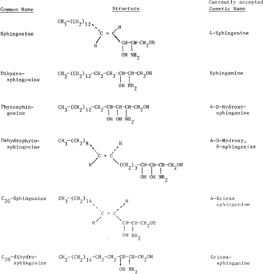
In the present system of nomenclature the C18 saturated base, dihydrosphingosine, is tentatively designated as sphinganine. According to this nomenclature, sphingosine is 4-sphingenine (the prefix 4 indicates the position of the double bond and phytosphingosine is termed 4-hydroxysphinganine. Homologues of C-18 are designated by an appropriate prefix (Table 1).
The primary amino group at C-2 in sphingosine is always N-acylated in sphingolipids, whereas the primary hydroxyl group at position 1 is either esterified or glycosylated. The N-acylated derivative of sphingosine, ceramide is the precursor of most of the sphingolipids and it has been isolated in the free state from neuronal and several other tissues (Gatt, 1963; Martensson, 1969; Samuelsson, 1969). Although various fatty acids have been detected in ceramide, the C20-C24 fatty acids predominate in neutral glycosphingolipids and sphingomyelin, whereas stearic acid is the major component of gangliosides.
CLASSIFICATION OF SPHINGOLIPIDS
The classification of sphingolipids is primarily based on the substituent groups attached to the hydroxyl group at C-1 of sphingosine or its derivatives. In phosphosphingolipids this hydroxyl group is esterified in a phosphate diester, as with phosphoryl choline in sphingomyelin, whereas in the glycosphingolipids the C-1 hydroxyl group is directly glycosylated by mono-, di-, or oligosaccharides. The glycosphingolipids acquire an anionic nature if the oligosaccharide moiety has acidic groups, as in sulfatide (galactose 3-sulphate) or in the gangliosides which contain sialic acid. Gangliosides are an important group of water-soluble acidic sphingolipids containing 3 or more hexose units attached to the C-1 hydroxyl of ceramide together with one or more sialic acid residues.
Sphingolipids are present in virtually all mammalian tissues and fluids although they are generally less abundant than the glycerides and cholesterol. They were once considered to be confined to the membranes of eukaryotic cells and to be absent from bacteria. Recent studies, however, have shown their occurrence in some of these organisms. In extra-neuronal tissues, the sphingolipids are believed to be localized mainly in plasma membrane and to contribute to the surface properties and to specific membrane functions of the cell.
CHEMICAL STRUCTURES AND OCCURRENCE
Structural studies of glycosphingolipids were mainly carried out in order to characterize the lipids accumulated in the brain and/or other tissues in inborn errors of metabolism such as Gaucher’s and Fabry’s disease. These diseases will be discussed in detail later in this volume. The structures of some glycosphingolipids are shown in Tables 2 and 3.
TABLE II
Structure of Some of the Neutral Sphingolipids
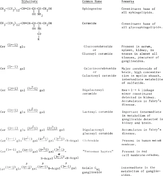
Cer = ceramide. glu = glucose. gal = galactose. N-Acgal = N-acetyl galactosamine.
TABLE III
Structure of Major Gangliosides and their Nomenclature
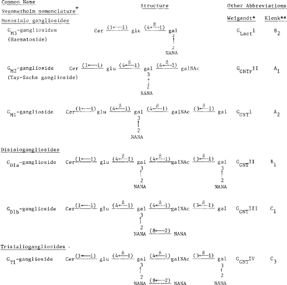
NANA = N-acetylneuraminic acid. Other abbreviations are same as in Table II.
+Svennerholm (1964)
*Kuhn & Weigandt (1963)
**Klenk & Gi...