
eBook - ePub
Immunology
An Illustrated Outline
David Male
This is a test
Partager le livre
- 152 pages
- English
- ePUB (adapté aux mobiles)
- Disponible sur iOS et Android
eBook - ePub
Immunology
An Illustrated Outline
David Male
Détails du livre
Aperçu du livre
Table des matières
Citations
À propos de ce livre
Immunology: An Illustrated Outline is both a guide to the essential principles of immunology and a concise dictionary of immunological terms. The book can be used to consolidate understanding in preparation for course exams and medical licensing exams, or as a refresher when immunology is encountered in related life sciences, such as microbiology, virology, and zoology.
The book is organized into five sections that represent the major topics in basic and clinical immunology. The Sixth Edition has been comprehensively revised to highlight the latest understanding of the field, particularly in the areas of innate immune defenses and antibody-based therapeutics.
- Concise explanations of immunological terms
- Full-color illustrations and micrographs to reinforce the text
- Each topic is set out in single- or double-page spreads
- Tables collate and summarize detailed information
Foire aux questions
Comment puis-je résilier mon abonnement ?
Il vous suffit de vous rendre dans la section compte dans paramètres et de cliquer sur « Résilier l’abonnement ». C’est aussi simple que cela ! Une fois que vous aurez résilié votre abonnement, il restera actif pour le reste de la période pour laquelle vous avez payé. Découvrez-en plus ici.
Puis-je / comment puis-je télécharger des livres ?
Pour le moment, tous nos livres en format ePub adaptés aux mobiles peuvent être téléchargés via l’application. La plupart de nos PDF sont également disponibles en téléchargement et les autres seront téléchargeables très prochainement. Découvrez-en plus ici.
Quelle est la différence entre les formules tarifaires ?
Les deux abonnements vous donnent un accès complet à la bibliothèque et à toutes les fonctionnalités de Perlego. Les seules différences sont les tarifs ainsi que la période d’abonnement : avec l’abonnement annuel, vous économiserez environ 30 % par rapport à 12 mois d’abonnement mensuel.
Qu’est-ce que Perlego ?
Nous sommes un service d’abonnement à des ouvrages universitaires en ligne, où vous pouvez accéder à toute une bibliothèque pour un prix inférieur à celui d’un seul livre par mois. Avec plus d’un million de livres sur plus de 1 000 sujets, nous avons ce qu’il vous faut ! Découvrez-en plus ici.
Prenez-vous en charge la synthèse vocale ?
Recherchez le symbole Écouter sur votre prochain livre pour voir si vous pouvez l’écouter. L’outil Écouter lit le texte à haute voix pour vous, en surlignant le passage qui est en cours de lecture. Vous pouvez le mettre sur pause, l’accélérer ou le ralentir. Découvrez-en plus ici.
Est-ce que Immunology est un PDF/ePUB en ligne ?
Oui, vous pouvez accéder à Immunology par David Male en format PDF et/ou ePUB ainsi qu’à d’autres livres populaires dans Medicina et Inmunología. Nous disposons de plus d’un million d’ouvrages à découvrir dans notre catalogue.
Informations
1 The Immune System
Introduction
The immune system has evolved to protect the body from damage caused by microorganisms—bacteria, fungi, viruses, and parasites. This defensive function is performed by leukocytes (white blood cells) and a number of accessory cells, which are distributed throughout the body but are found particularly in lymphoid organs, including the bone marrow, thymus, spleen, and mucosa-associated lymphoid tissues (MALT). Lymphoid organs are strategically placed to protect different areas of the body from infection. Cells migrate between these tissues via the bloodstream and lymphatic system. As they do so they interact with each other to generate coordinated immune responses, aimed at eliminating pathogens or minimizing the damage they cause.
Lymphocytes are key cells controlling the immune response. They do so by recognizing molecules produced by pathogens. They can also recognize molecules on the cells of the body, although they do not normally react against the body's own tissues. Molecules recognized by lymphocytes are referred to as “antigens.” Lymphocytes are of two main types: B cells, which produce antibodies, and T cells, which have a number of functions, including (1) helping B cells to make antibodies; (2) recognizing and destroying cells that have become infected with intracellular pathogens; (3) activating phagocytes to destroy pathogens that they have taken up, and (4) regulating the level and quality of the immune response. Lymphocytes recognize foreign material by specific cell-surface antigen receptors. To recognize the enormous variety of different molecules, the antigen receptors must be equally diverse. Each lymphocyte makes only one type of antigen receptor and thus can only recognize a very limited number of antigens, but as the receptors differ on each clone of cells, the lymphocyte population, as a whole, can recognize a vast range of different antigens. A third population of lymphocytes, the innate lymphoid cells (ILCs) do not have specific antigen receptors but generate a variety of immune defense functions.
Phagocytes include blood monocytes, macrophages, and neutrophils. They can internalize (phagocytose) pathogens, antigens, and cell debris and break them down. Antibodies and various other immune recognition molecules bound to the pathogens facilitate this process. Macrophages can also process and present antigens, so that they can be recognized by T cells.
Accessory cells include eosinophil and basophil granulocytes, mast cells, platelets, and antigen-presenting cells (APCs). Eosinophils have a role in protection against some parasites. Basophils, mast cells, and platelets contain a variety of molecules that mediate inflammation. APCs are a functionally defined group of cells; both B cells and macrophages can present antigen, but leukocyte dendritic cells are particularly important in presenting antigen to naive T cells, which have not previously encountered their specific antigen.
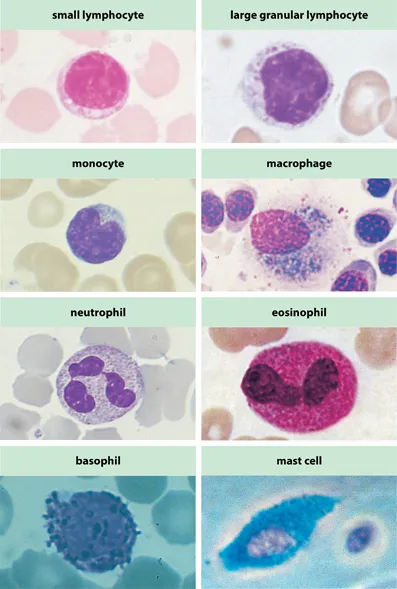
Fig. 1.1 Cells involved in the immune response.
Macrophage courtesy of A. V. Hoffbrand.
Lymphocytes
Lymphocytes constitute about 20% of the total blood leukocytes. The two major populations of lymphocytes, T cells and B cells, are small lymphocytes responsible for recognizing antigens or antigen fragments. Innate lymphoid cells (ILCs) are a heterogenous group, which can carry out many of the functions of T cells—the group includes natural killer (NK) cells.
Large granular lymphocytes (LGLs) are morphologically defined cells containing large amounts of cytoplasm, with azurophilic granules, which constitute 5–15% of the blood T cells. Both NK cells and γδ T cells have this morphology.
T cells are lymphocytes that develop in the thymus. This organ is seeded by lymphocytic stem cells from the bone marrow during embryonic development. The cells then develop their T-cell antigen receptors (TCR) and differentiate into the two major peripheral T-cell subsets; the helper T cells express CD4, and the cytotoxic T cells express CD8. T cells can also be differentiated into two populations according to whether they use an aβ (TCR2) or a γδ (TCR1) antigen receptor. The essential role of T cells is to recognize antigens associated with cells of the host.
γδ T cells express the γδ form of the T-cell receptor. They constitute <5% of the total T cells, but they are more common in particular sites, including the gut, skin, and vagina. They branch early from the main thymic developmental pathway and recognize different antigens from αβ T cells, including carbohydrates and intact proteins.
Intraepithelial lymphocytes (IELs) are mixed populations of cells found in submucosal tissues. A total of 10–40% are γδ T cells, with a dendritic appearance. The remainder are mostly CD8+ T cells.
Cytotoxic T lymphocytes (CTLs/TC) are capable of destroying virally infected or allogeneic cells. Most CTL cells express CD8 and recognize antigen associated with major histocompatibility complex (MHC) class I molecules, which may be expressed on all nucleated cells of the body.
Helper T (TH) cells perform a number of functions, including helping B cells to divide, differentiate, and secrete antibody, activating macrophages to destroy pathogens that they have phagocytosed and recruiting cells to sites of inflammation. The functions are carried out by different subsets of TH cells, which differentiate from a common precursor (TH0) and can be distinguished by the cytokines, which they secrete. The majority of TH cells express CD4 and recognize antigenic peptides presented on the surface of APCs by MHC class II molecules.
TH1, TH2, and TH17 cells are subsets of helper T cells originally identified according to the cytokines they produce. Differentiation of TH1 cells is promoted by interleukin-12 (IL-12) and interferon-γ (IFNγ), TH2 cells by IL-4, and TH17 cells by transforming growth factor-β (TGFβ) and IL-6. Dendritic cells are antigen-presenting cells, which are most effective at presenting antigen to naive T cells. TH1 cells can recognize antigen presented by mononuclear phagocytes, and they interact with these cells by releasing IFNγ, which acts as a macrophage activation factor. TH2 cells release cytokines, such as IL-4 and IL-5, which are required for B-cell development into plasma cells. Both TH1 and TH2 cells can modulate the antibody response by affecting the classes of immunoglobulin produced. TH17 cells release cytokines that promote inflammatory responses, particularly by acting on neutrophils. Some cell-surface markers are preferentially expressed on a subset of the helper T cells. For example, the chemokine receptors CCR5 and CXCR3 are more prevalent on TH1 cells, whereas CCR3 and CCR4 are higher on TH2 cells. All helper T cells can promote the development and activation of cytotoxic T cells and NK cells, which recognize and kill infected target cells.
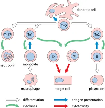
Fig. 1.2 Lymphocyte interactions.
Regulatory T cells (TREG), identified by the expression of the transcription factor Foxp3 and/or high expression of the IL-2 receptor (CD25), constitute 5–10% of the total T cells. They can be either CD4+ or CD8+ and are important in controlling secondary immune responses and inflammation, particularly in the gut. They also limit some autoimmune and hypersensitivity reactions, acting by direct cell-cell interactions, or by the release of anti-inflammatory cytokines including IL-10, IL-35, and TGFβ. They can also inhibit activation of other T cells by mopping up IL-2, which is required for T-cell proliferation.
B cells are lymphocytes that develop in the fetal liver and subsequently in bone marrow. In birds, they develop in a specialized organ, the bursa of Fabricius. Mature B cells express surface immunoglobulin, which acts as the B-cell antigen receptor (BCR). They are distributed throughout the secondary lymphoid tissues, particularly in the follicles of lymph nodes, spleen, and Peyer's patches. They respond to antigenic stimuli by dividing and differentiating into plasma cells.
Plasma cells/Antibody-forming cells (AFCS) are terminally differentiated B cells, with expanded cytoplasm containing arrays of rough endoplasmic reticulum, devoted to the synthesis of secreted antibody. Plasma cells are seen in the red pulp of spleen, the medulla of lymph nodes, the MALT, and occasionally in sites of inflammation.
B1 and B2 cells are B-cell subsets. In adults most B cells are of the B2 subset. They generate a wide range of antigen receptors, mature in germinal centers and respond well to T-dependent antigens and costimulation via CD40. The B1 subset was originally distinguished by the phenotyp...