2Diagnosis
Clinical features
The classic presentation of BPDCN (in more than 70% of cases) is the clinical leukemic features of hematodermic lesions with bone marrow involvement. Every time physicians see such a patient, a possible diagnosis of BPDCN should come to mind.
Most patients with BPDCN are in a good general state of health at diagnosis,1–3 but the following clinical features may be present.
Cutaneous lesions are the hallmark of BPDCN, and in 85–90% of cases they are the first lesion to appear, even when the diagnosis is made retrospectively. Dermatologists are often the first specialists confronted with the disease.4 The lesions are heterogeneous, ranging from maculonodular lesions to dystrophic xanthomatosis. They may be unique or diffuse and largely disseminated. The elementary lesion is frequently infiltrated and presents in the form of a violaceous plaque or nodule, frequently spreading to the entire skin. Sometimes, lesions may appear hemorrhagic or ecchymotic with a purpuric aspect (petechiae), suggestive of problems with hemostasis, like platelet disorders or von Willebrand disease. Pruritus is present only exceptionally. Sometimes lesions may be painful, with an inflammatory aspect. Bruise-like patches disseminated on the upper side of the body appear to be the most common presenting sign of BPDCN on the skin.1,4–8 Figure 2.1 illustrates various skin lesions observed in patients with BPDCN.
To evaluate the dermatologic involvement, it is important to use a standardized score like the one dermatologists use to assess hematodermic malignancies.4 We recommend the use of the modified Severity-Weighted Assessment Tool (mSWAT).9,10 This is mandatory in therapeutic research protocols and very useful in clinical practice for effective follow-up of patients. First, the extent of the total body surface area (BSA) affected by each lesion type (patch, plaque and tumor or ulceration) is recorded on a body map (Figure 2.2). A 1-cm grid is then randomly placed on the drawing, and the number of grid intersections overlying each type of lesion is counted. The mean of six grid placements and counts is divided by the maximum possible number of intersections for the body diagram and multiplied by 100 to give the percentage total BSA involvement for each lesion type. Each total is then multiplied by a severity-weighting factor. The mSWAT score is the sum of these three totals on a scale of 0 to 400: (patch %TBSA × 1) + (plaque %TBSA × 2) + (tumor or ulcer %TBSA × 4) (Table 2.1).
Figure 2.1 Heterogeneous cutaneous lesions are the hallmark of BPDCN. Note that the elementary lesion presents as a violaceous plaque or nodule. Photographs courtesy of Drs Andrew Lane, Vincent Marmouset and Bérengère Gruson. Middle row images reproduced with permission from Pemmaraju et al. 2019.11
Figure 2.2 mSWAT body map for a hypothetical patient with BPDCN. Patch disease is drawn as single-hatched, plaque disease as cross-hatched, and tumors and ulceration as solid shaded areas. A point-counting grid is used to calculate the total BSA of involvement for each lesion type from which the total mSWAT score is calculated (see text and Table 2.1).
TABLE 2.1
Calculating the mSWAT score
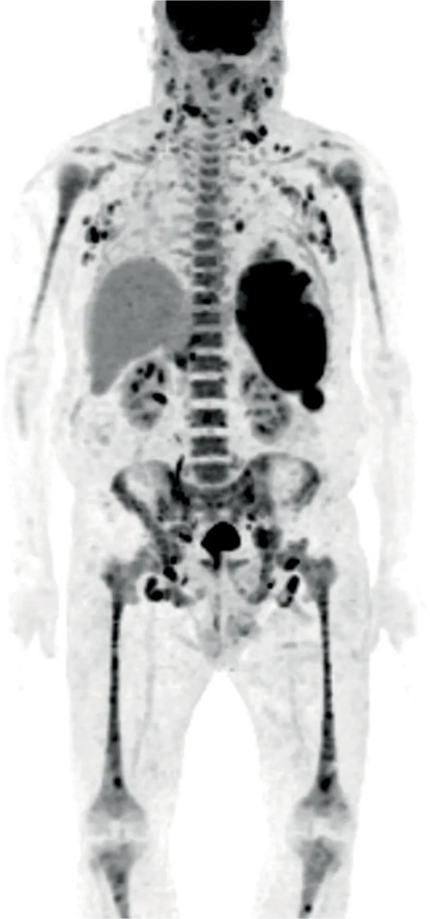
Extracutaneous manifestations of BPDCN are less frequent (Figure 2.3). Lymphadenopathies are present in 40% of cases and hepatosplenomegaly in 20%. Visceral extranodal involvement is rare. Specific lesions have been reported, for example, in the lungs, digestive tract, urinary tract, breasts and paranasal sinuses, but virtually all organs may be involved.1,2,5,6
The involvement of other organs has no specific characteristics and is clinically perceptible or easily evaluated with CT or PET scans at the initial evaluation when patients are suspected of having BPDCN (Figure 2.4). PET is a good tool for the initial diagnostic staging of the disease, as it can then provide a useful evaluation of tumoral response thereafter.2,12
Neurological involvement in BPDCN warrants special attention. As in other forms of leukemia, involvement of the cerebrospinal fluid (CSF) is present in up to 10% of patients at diagnosis and appears to be more frequent after relapse (20%).5,13,14 A retrospective study of 109 patients found three cases with CSF involvement, even in the absence of specific symptoms, and two cases with ocular infiltration.5 This raises the question as to whether specific CNS investigation and/or prophylaxis is needed in all patients diagnosed with BPDCN, especially in this era of new targeted molecules and immunotherapies.
Figure 2.3 Organ involvement in the clinical presentation of BPDCN.
Figure 2.4 PET-CT scan of a patient with BPDCN at diagnosis, showing extensive organ involvement.
Overt leukemia is the initial presentation in 10% of patients, resulting in cytopenia (particularly thrombocytopenia in 75% of cases), fatigue, bleeding, bruising and fever, but no cutaneous lesions.
Cytomorphological features
The diagnosis of BPDCN can only be established after histopathological assessment of the involved tissue (skin or bone marrow) with the use of specific immunophenotypic markers, or by evaluating blast morphology plus immunophenotyping of infiltrated blood and/or bone marrow.
Complete blood cell count (CBC) at diagnosis yields variable findings in patients with BPDCN. These can range from absence of cytopenia and no blasts on blood smear (if the diagnosis is made by a dermatologist on isolated cutaneous lesions), to frequent cytopenia, due to bone marrow replacement, and blasts in the peripheral blood (in two-thirds of patients). The latter more advanced pathology is usually diagnosed by a hematologist. However, hyperleukocytosis with a high number of blasts in the blood is uncommon. It is noteworthy that myelemia was found to be associated with poor prognosis in a large series of patients with BPDCN.5 If there is a primary history of another hemopathy, the CBC can harbor monocytosis (if BPDCN arose from CMML), dysmorphia and macrocytosis (if a myelodysplasia is present).
Immunohistochemistry. The skin is one of the most common initial biopsy sites in BPDCN. Neoplastic cells usually infiltrate the superficial and/or deep dermis in a dense diffuse or nodular growth pattern, usually with sparing of adnexal structures (Figure 2.5). Cases with micronodular dermal infiltration and perivascular/periadnexal involvement are less common, as are occasional cases with interstitial dermal infiltration. While subcutaneous extension is common in BPDCN, a grenz zone is typically present and epidermotropism is very uncommon. In the skin, as in other tissue samples, BPDCN usually consists of a monotonous population of neoplastic cells with scant or moderate amounts of cytoplasm, round nuclei with fine (blastoid) chromatin and inconspicuous nucleoli. However, nuclear morphology in BPDCN is variable and can include cases with larger nuclei and prominent nucleoli (immunoblastoid) and others with small nuclei characterized by vesicular or condensed chromatin (lymphoma-like). Mitotic figures and apoptotic bodies are common, but tumor necrosis is variable.
Figure 2.5 BPDCN involving the skin. (a) The neoplastic cells are round and monotonous, and infiltrate the skin in a diffuse and nodular pattern with epidermal and adnexal sparing. 100× magnification; hematoxylin and eosin. The neoplastic cells are positive by immunohistochemistry for the following markers: (b) CD4; (c) CD56; (d) CD123. 200× magnification; hematoxylin counterstain.
These characteristics are also observed in other organs. In the lymph nodes, the neoplastic infiltrate is usually interfollicular in distribution. Bone marrow involvement is diffuse in most patients who have a leukemic presentation. In those who have more limited bone marrow involvement, BPDCN often exhibits an interstitial growth pattern.
Blast morphology. Blastic infiltration is most easily discernible on bone marrow aspiration, but may be absent if the pathology is only cutaneous. It should be noted that the morphological features of the bl...