Applications to Dosimetry, Imaging, and Preclinical Radiotherapy
Joao Seco, Frank Verhaegen, Joao Seco, Frank Verhaegen
This is a test
This is a test
Partager le livre
274 pages
English
ePUB (adapté aux mobiles)
Disponible sur iOS et Android
eBook - ePub
Monte Carlo Techniques in Radiation Therapy
Applications to Dosimetry, Imaging, and Preclinical Radiotherapy
Joao Seco, Frank Verhaegen, Joao Seco, Frank Verhaegen
Détails du livre
Aperçu du livre
Table des matières
Citations
À propos de ce livre
Thoroughly updated throughout, this second edition of Monte Carlo Techniques in Radiation Therapy: Applications to Dosimetry, Imaging, and Preclinical Radiotherapy, edited by Joao Seco and Frank Verhaegen, explores the use of Monte Carlo methods for modelling various features of internal and external radiation sources.
Monte Carlo methods have been heavily used in the field of radiation therapy in applications such as dosimetry, imaging, radiation chemistry, modelling of small animal irradiation units, etc. The aim of this book is to provide a compendium of the Monte Carlo methods that are commonly used in radiation therapy applications, which will allow students, postdoctoral fellows, and university professors to learn and teach Monte Carlo techniques. This book provides concise but detailed information about many Monte Carlo applications that cannot be found in any other didactic or scientific book.
This second edition contains many new chapters on topics such as:
Monte Carlo studies of prompt gamma emission
Developments in proton imaging
Monte Carlo for cone beam CT imaging
Monte Carlo modelling of proton beams for small animal irradiation
Monte Carlo studies of microbeam radiation therapy
Monte Carlo in micro- and nano-dosimetry
GPU-based fast Monte Carlo simulations for radiotherapy
This book is primarily aimed at students and scientists wishing to learn and improve their knowledge of Monte Carlo methods in radiation therapy.
Foire aux questions
Comment puis-je résilier mon abonnement ?
Il vous suffit de vous rendre dans la section compte dans paramètres et de cliquer sur « Résilier l’abonnement ». C’est aussi simple que cela ! Une fois que vous aurez résilié votre abonnement, il restera actif pour le reste de la période pour laquelle vous avez payé. Découvrez-en plus ici.
Puis-je / comment puis-je télécharger des livres ?
Pour le moment, tous nos livres en format ePub adaptés aux mobiles peuvent être téléchargés via l’application. La plupart de nos PDF sont également disponibles en téléchargement et les autres seront téléchargeables très prochainement. Découvrez-en plus ici.
Quelle est la différence entre les formules tarifaires ?
Les deux abonnements vous donnent un accès complet à la bibliothèque et à toutes les fonctionnalités de Perlego. Les seules différences sont les tarifs ainsi que la période d’abonnement : avec l’abonnement annuel, vous économiserez environ 30 % par rapport à 12 mois d’abonnement mensuel.
Qu’est-ce que Perlego ?
Nous sommes un service d’abonnement à des ouvrages universitaires en ligne, où vous pouvez accéder à toute une bibliothèque pour un prix inférieur à celui d’un seul livre par mois. Avec plus d’un million de livres sur plus de 1 000 sujets, nous avons ce qu’il vous faut ! Découvrez-en plus ici.
Prenez-vous en charge la synthèse vocale ?
Recherchez le symbole Écouter sur votre prochain livre pour voir si vous pouvez l’écouter. L’outil Écouter lit le texte à haute voix pour vous, en surlignant le passage qui est en cours de lecture. Vous pouvez le mettre sur pause, l’accélérer ou le ralentir. Découvrez-en plus ici.
Est-ce que Monte Carlo Techniques in Radiation Therapy est un PDF/ePUB en ligne ?
Oui, vous pouvez accéder à Monte Carlo Techniques in Radiation Therapy par Joao Seco, Frank Verhaegen, Joao Seco, Frank Verhaegen en format PDF et/ou ePUB ainsi qu’à d’autres livres populaires dans Medicina et Oncologia. Nous disposons de plus d’un million d’ouvrages à découvrir dans notre catalogue.
10Monte Carlo Studies for Microbeam Radiation Therapy
Susanna Guatelli
Centre For Medical and Radiation Physics, University of Wollongong
Stefan Bartzsch
Department of Radiation Oncology, School of Medicine and Klinikum rechts der Isar, Technical University of Munich (TUM)
Matthew Cameron
Centre For Medical and Radiation Physics, University of Wollongong, and ANSTO-Australian Synchrotron
Michael L. F. Lerch
Centre For Medical and Radiation Physics, University of Wollongong
Jason Paino
Centre For Medical and Radiation Physics, University of Wollongong
DOI: 10.1201/9781003212485-14
10.1 Introduction
10.2 Fundamental Physics and Technology Concepts of MRT
Generation of Synchrotron Radiation
10.3 Physics Processes Important for MRT Dosimetry
10.4 Validation of Monte Carlo Simulations for MRT
10.5 Modelling the Target Treatment
10.6 Monte Carlo-Based MRT Treatment Planning
Conversion of CTs, Organs with Microstructure
Monte Carlo Dosimetric Engines for MRT Treatment Planning
10.7 Conclusions
Acknowledgments
References
10.1 Introduction
Microbeam radiation therapy (MRT) is a radiotherapy modality that uses arrays of highly collimated, quasi-parallel micrometer-sized X-ray beams to deliver high radiation doses (Bravin et al. 2015). Its inception dates back to the late 1950s when H. J. Curtis and C. P. Baker, Brookhaven National Laboratory (BNL), the United States, discovered that the threshold dose for normal tissue damage was several hundred Grays when irradiating a mouse brain with deuteron microbeams (Zeman et al. 1959, 1961). It was then found that the tolerance of normal tissue to radiation dose increases dramatically, which is inversely proportional to the size of the radiation field (Hopewell et al. 1987). This phenomenon is known as the dose–volume effect.
Although the microbeam dose–volume effect was discovered in the 1950s, the investigation of the use of multiple parallel planar X-ray microbeams for radiotherapy started only in the 1990s by D. Slatkin and colleagues at the BNL (Slatkin et al. 1992 and 1995, Laissue et al. 1998, 1999) when it became technologically possible to generate high-intensity X-ray beams with a very low divergence. Despite the encouraging results, the program was interrupted at the BNL and was started at the European Synchrotron Radiation Facility (ESRF), in Grenoble, France, in the mid-1990s (Bravin et al. 2015), and it is still active. In addition to the ESRF, MRT has been subject of research at SPring-8 (Hyogo Prefecture, Japan), the Australian Synchrotron (AS) (Melbourne, Australia), and the Canadian Light Source (Saskatoon, Canada) (Bartzsch et al. 2020), attracting the attention of several research groups internationally (Bravin et al. 2015).
Research performed in the past 25 years demonstrates that MRT delivered at the third-generation synchrotron facilities is particularly promising for patients with malignant central nervous system tumors, for whom there is currently no satisfactory therapy available (Grotzer et al. 2015). MRT treatment can be seen as a combination of spatially fractionated X-ray beam delivery, coupled with high dose rates (up to the order of 106 Gy/s) typical of FLASH therapy (Wilson et al. 2020), and current research is focused in translating MRT from a preclinical to a clinical stage (Engels et al. 2020).
The first Monte Carlo (MC) simulations for MRT were performed by Slatkin et al. (1992) using an early version of EGS4 (INHOM) (Nelson et al. 1985). Since then, all the most popular general-purpose MC codes describing particle transport and interactions in matter, Geant4 (Agostinelli et al. 2003, Allison et al. 2006, 2016), PENELOPE (Sempau et al. 1997, 2011, Salvat et al. 2008), EGS4 (Nelson et al. 1985), EGS5 (Hirayama et al. 2005), EGSnrc (Mainegra-Hing et al. 2020), MCNP6 (Goorley et al. 2013), and MCNPX (McKinney et al. 2006), have been used in MRT-related research to characterize MRT beamlines, to perform dosimetric calculations in the treatment target, and to investigate novel detector technology for MRT quality assurance (Bravin et al. 2015, Bartzsch et al. 2020, Brauer-Krisch et al. 2015).
After a brief description of the fundamental physics concepts and technology involved with MRT, this chapter will focus on important aspects to consider in MC simulations aimed to model MRT beamlines and to perform dosimetric calculations in the treatment target. We will refer to the specific case of the MRT research beamline of the AS as example.
We will then describe the state-of-the-art research aimed to develop MRT Treatment Planning System (TPS) engines based on MC simulations. Finally, we will conclude with some remarks on possible next development of MC simulations for MRT.
10.2 Fundamental Physics and Technology Concepts of MRT
With respect to conventional X-ray radiotherapy, the X-ray beam for MRT is characterized by a very small divergence and a very high dose rate (from hundreds to thousands of Gy/s). The generation of a X-ray field with these features is currently only feasible at the third-generation synchrotron facilities.
In this section, we will describe briefly the beamline of the AS used for MRT research. For more details, the reader is referred to Stevenson et al. (2017a, 2017b) and Livingstone et al. (2017).
At the AS, the injection system accelerates electrons to a highly relativistic speed, reaching an energy of 3 GeV (Friis-Nielsen et al. 2006). Then, the electron bunches are extracted and injected in the storage ring (storage ring current = 200 mA, data from Livingstone et al. 2017).
The Imaging and Medical Beamline (IMBL), sketched in Figure 10.1, is one of ten beamlines at the AS and is dedicated to medical research.
FIGURE10.1 Schematic of the Imaging and Medical beamline (IMBL), Australian Synchrotron. The path of the electron bunches is represented by the red track, while the synchrotron radiation, produced in the wiggler, by the blue beam.
Once the electron bunches are extracted from the AS storage ring and injected in the IMBL, they traverse a superconducting multipole wiggler consisting of 30 pole pairs with 52 mm period.
The magnetic field in the wiggler can be described with a sinusoidal curve as follows: where z is the distance along the wiggler axis and λu is the magnetic period (Kim 2020). The standard operating magnetic field B0 of the wiggler is 3 T (Livingstone et al. 2017); however, also other magnitudes (e.g., 1.4, 2 and 4 T) are available. In reality, the magnetic field within the wiggler is not a simple sinusoid due to the effect of boundary conditions at the ends of the wiggler, interaction between adjacent magnetic poles, and magnetic field nonuniformities. Nevertheless, this is the approximation which is usually used (Tanaka 2014).
Because of the magnetic field in the wiggler, the electrons “zig-zag” in their path and, because of the trajectory bending, originate synchrotron radiation as shown in Figure 10.2. This process is described in more detail in Section 10.2.1.
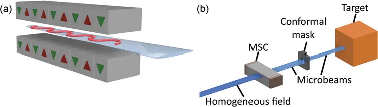
FIGURE10.2 (a) Principle of generation of synchrotron radiation generation by means of a high-energy electron traversing a wiggler. The red track represents the trajectory of the electron, while the blue beam represents the emerging synchrotron radiation. The red and green arrows indicate the inverse polarity of the magnets in the wiggler. (b) Principle of X-ray microbeam array generation. A multislit collimator (MSC) shapes microbeams from a X-ray broadbeam.
After traversing the wiggler, the electrons are diverted by a bending magnet and the synchrotron radiation, which is highly polarized in the electron orbital plane (see more details in Section 10.2.1), is transported through the beamline.
When generated, the synchrotron radiation has a continuous spectrum spanning from the infrared to the X-ray region. Permanent filters (sketched in Figure 10.1) of graphene and high-density graphite absorb low-energy photons and avoid excessive thermal load on beamline components (Livingstone et al. 2017). Additional filtration, which can be used at the IMBL, consists of one of the following combinations: Cu/Cu, Cu/Al, Al/Cu (or Cu/Al), Al/Al, AlMo/AlMo (Stevenson et al. 2017b). In this chapter, as practical example, we will refer to the Cu/Cu filtration, consisting of two copper paddles, each 2 mm thick, placed at 45° to the incident beam (Stevenson et al. 2017b).
An increase in the magnetic field (B0) translates in an increase of the photon flux and in the hardening of the spectrum. For example, using the same Cu/Cu filtration, the weighted mean value of the energy spectrum increases from 71 to about 95 keV when increasing B0 from 2 to 3 T (Dipuglia et al. 2019).