![]()
Indications for Laser Applications
Bogdan Allemann I, Goldberg DJ (eds): Basics in Dermatological Laser Applications.
Curr Probl Dermatol. Basel, Karger, 2011, vol 42, pp 81-96
______________________
Benign Pigmented Lesions
Inja Bogdan Allemanna · David J. Goldbergb,c
aKlinik für Plastische Chirurgie und Handchirurgie, Universitätsspital Zürich, Zürich, Switzerland; bSkin Laser & Surgery Specialists of New York & New Jersey, andcMount Sinai School of Medicine, New York, N.Y., USA
______________________
Abstract
Benign pigmented lesions are a frequent complaint in dermatological patients, especially those seeking advice and therapy in a laser or cosmetic practice. Significant advances in laser technology over the last decades now allow us to effectively and safely treat various benign pigmented lesions. However, a thorough understanding of the biology of the lesion to be treated, the physical properties of the lasers to be used, and laser-tissue interactions is crucial for a successful and safe treatment. This chapter will give an overview of the types of benign pigmented lesions that can be treated with lasers and the specific lasers used to treat them.
Copyright © 2011 S. Karger AG, Basel
Our skin color is determined by the activity of our pigment-producing melanocytes. The distribution of melanin varies from skin phototype to skin phenotype. Individuals with darker skin types have the same amount of melanocytes as lighter-skinned people; however, they have larger melanosomes that contain more melanin pigment. These melanosomes are also more evenly dispersed throughout the cytoplasm.
Melanin production takes place within the melanosome, which is an intracytoplasmic organelle within the melanocyte. The enzyme tyrosinase is the key enzyme in the biosynthesis of melanin. Melanocytes are found in the basal layer of the epidermis. In normal skin, roughly every tenth cell in the basal layer is a melanocyte. With its dendrites, one melanocyte can reach as far as the stratum spinosum, contacting roughly 30-40 keratinocytes, which is called an epidermal melanin unit. The melanocyte interacts with the keratinocytes by transferring melanosomes from the dendrites of the melanocyte into neighboring keratinocytes.
Pigmented lesions show either an increase in melanin production with excessive melanin deposition in melanosomes or an increase in the density of active melanocytes.
Selective Photothermolysis
The principle of selective photothermolysis first described by Anderson and Parrish [1] more than 2 decades ago led to a revolution in the treatment of pigmented lesions. As described in chapter explaining the physics of lasers in more detail [Bogdan Allemann and Kaufman, pp. 7-23], the principle states firstly that a target chromophore can selectively be destroyed, sparing the surrounding tissue from damage, by choosing the wavelength that corresponds to the absorption maximum/spectrum of that target chromophore. Secondly, by adapting the pulse width to be equal to or shorter than the thermal relaxation time (TRT, the time needed by a structure to cool down to half the temperature to which it was heated) of the target chromophore, heat is selectively generated in the target and does not harm the surrounding tissue. Finally, the energy of the laser pulse must be high enough to actually destroy the target chromophores. These three parameters, wavelength, pulse duration and energy have to be chosen with respect to the target in order to selectively destroy it and leave surrounding tissue unharmed [2-4].
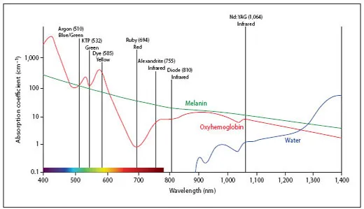
Fig. 1. Absorption spectrum of melanin.
Physical Principles in the Treatment of Pigmented Lesions
The target chromophore in the treatment of pigmented lesions is the melanin-containing melanosome. Experimental studies have demonstrated that Q-switched (QS) lasers can target and selectively destroy melanosomes [5, 6].
Hence, the three parameters defining selective destruction by the principle of selective photothermolysis - namely wavelength, pulse duration and fluence - have to be chosen according to the characteristics of the melanosome.
Wavelength
The absorption spectrum of melanin is broad and ranges from 351 to 1,064 nm, with its maximum in the ultraviolet region, a gradual decrease throughout the visible region, and an extension into the near-infrared region [7] (fig. 1). Therefore, the wavelength of the chosen laser should correspond to the absorption spectrum of melanin, with an optimum between 600 and 1,100 nm, as light within these wavelengths is able to penetrate into the dermis without significant absorption by competing chromophores, such as hemoglobin and water [37].
The exact choice of the laser wavelength used depends upon the depth of the pigmented lesion as well as on the patient's skin type. Within the visible range, shorter wavelengths penetrate less deeply but show stronger melanin absorption, while long wavelengths penetrate deep into the tissue due to decreased scatter but have poorer melanin absorption; hence, treatment efficacy decreases with increasing wavelength [8-10]. Consequently, epidermal pigment can be targeted with wavelengths that penetrate more superficially, while dermal pigment needs to be targeted with wavelengths that can successfully penetrate deeply into the tissue.
However, as the laser light does not differentiate between pathological (i.e., excess pigment) and normal melanin, defining the patients’ skin type, special care should be taken when treating darker skin types. Lasers with high melanin absorption clearly carry an increased high risk of side effects, such as pigment alterations, and should thus be avoided in darker-skinned patients. Patients with Fitzpatrick skin phototype >IV are safest treated with longer wavelengths, such as the 1,064 nm Nd:YAG laser, in order to prevent too much absorption of epidermal melanin and consecutive pigmentary alterations.
Pulse Duration
Melanin is found within the melanosomes. Melanosomes are very small organelles, measuring roughly 0.5-1 μm, and consequently have a short TRT. The TRT is directly proportional to the square of the size of the target; thus, for melanosomes it lies between 0.25 and 1 μs. Consequently, lasers with pulses in the nanosecond range, as is the case with QS lasers, should be used in order to confine the heat within the target and not damage surrounding tissue.
Fluence
Wavelength and pulse duration are both important factors in treating pigmented lesions. Fluence (energy delivered over treated area) is also a highly important treatment parameter. A minimal fluence is required for treatment success in epidermal pigmented lesions. Although at the requisite fluence, epidermal pigmented lesions can be treated with a small treatment spot size, dermal pigmented lesions are different. With dermal pigmented lesions, the larger the delivered spot size, the greater the chances of success at every chosen fluence.
Spot Size
When treating pigmented lesions, the selection of the spot size, as described above, is also crucial. Although a larger spot size decreases peripheral scatter and, subsequently, results in a higher percentage of energy being delivered to the target, the spot size should not be larger than the lesion treated in order to prevent pigmentary changes to the surrounding skin. This is especially crucial when treating darker-skinned patients. The spot size should thus be chosen according to the size of the targeted structure. Ideally, the largest spot size that accommodates the treated lesion should be chosen.
Lasers Used in the treatment of Pigmented Lesions
When treating pigmented lesions, 4 categories of lasers or light devices can be used: (1) the pigment-selective QS lasers, (2) the less-selective long-pulsed (lp) lasers and intense pulsed light (IPL), (3) the non-pigment-specific ablative lasers, and (4) the recently described non-pigment-specific fractionated lasers [11].
Based on the concept of selective photother-molysis, the treatment of pigmented lesions should ideally be performed with lasers that are able to emit high-energy pulses within the nanosecond range. These lasers are referred to as QS lasers. The term ‘Q-switching’ refers to a switch resulting in the release of all the energy in one short powerful pulse, allowing for selective photothermolysis [12]. The method of Q-switching allows one to produce very short laser pulses in the nanosecond domain, in order to treat small micrometer-sized targets, such as melanin. QS lasers currently on the market are the QS ruby at 694 nm, the QS alexandrite at 755 nm, the QS Nd:YAG at 1,064 nm and the frequency-doubled QS Nd:YAG at 532 nm.
The less-pigment-selective lp lasers used to treat pigmented lesions are the lp ruby at 694 nm, the lp alexandrite at 755 nm, the lp Nd:YAG at 1,064 nm and the diode laser at 800 nm. Due to their pulse durations within the millisecond range, which exceed the TRT of the target chromophore melanin, lp lasers bear the risk of causing damage to the surrounding tissue, which can result in scarring. Therefore, the energy chosen must be low. Lp lasers can better treat epidermal pigmented lesions than dermal ones, due to the thermal damage they cause to melanosome-containing keratinocytes. Dermal lesions should not be treated with lp lasers because of the associated higher risk of scarring [13]. It should be noted, however, that lp lasers are clearly less effective than pigment-specific QS lasers, are associated with a higher risk of complications, and require more treatments to achieve optimal clearing. With pulse durations in the millisecond range, the lp lasers are however ideal to treat hair, where the target chromophore still is melanin, but longer pulse durations are necessary to achieve destruction of the hair follicle. This will be explained in greater detail in the chapter on hair removal [Haedersdal and Haak, pp. 111-121].
Polychromatic light generated by IPL sources (515-1,200 nm) can also be used to treat epidermal pigmented lesions. Due to the polychromatic nature of the IPL devices, competing chromophores, such as hemoglobin, need to be taken into account. In order to avoid absorption by hemoglobin, compression may be us...