Biological Sciences
Haemoglobin
Haemoglobin is a protein found in red blood cells that is responsible for transporting oxygen from the lungs to the body's tissues and returning carbon dioxide from the tissues back to the lungs. It consists of four subunits, each containing a heme group with an iron atom that binds to oxygen. This crucial role in oxygen transport makes haemoglobin essential for sustaining life.
Written by Perlego with AI-assistance
Related key terms
1 of 5
9 Key excerpts on "Haemoglobin"
 No longer available |Learn more
- (Author)
- 2014(Publication Date)
- Orange Apple(Publisher)
The molecule also carries the important regulatory molecule nitric oxide bound to a globin protein thiol group, releasing it at the same time as oxygen. Hemoglobin is also found outside red blood cells and their progenitor lines. Other cells that contain hemoglobin include the A9 dopaminergic neurons in the substantia nigra, macrophages, alveolar cells, and mesangial cells in the kidney. In these tissues, hemoglobin has a non-oxygen-carrying function as an antioxidant and a regulator of iron metabolism. Hemoglobin and hemoglobin-like molecules are also found in many invertebrates, fungi, and plants. In these organisms, hemoglobins may carry oxygen, or they may act to transport and regulate other things such as carbon dioxide, nitric oxide, hydrogen sulfide and sulfide. A variant of the molecule, called leghemoglobin, is used to scavenge oxygen, to keep it from poisoning anaerobic systems, such as nitrogen-fixing nodules of leguminous plants. ________________________ WORLD TECHNOLOGIES ________________________ Research history The oxygen-carrying protein hemoglobin was discovered by Hünefeld in 1840. In 1851, Otto Funke published a series of articles in which he described growing hemoglobin crystals by successively diluting red blood cells with a solvent such as pure water, alcohol or ether, followed by slow evaporation of the solvent from the resulting protein solution. Hemoglobin's reversible oxygenation was described a few years later by Felix Hoppe-Seyler. In 1959 Max Perutz determined the molecular structure of hemoglobin by X-ray crystallography. This work resulted in his sharing with John Kendrew the 1962 Nobel Prize in Chemistry. The role of hemoglobin in the blood was elucidated by physiologist Claude Bernard. The name hemoglobin is derived from the words heme and globin , reflecting the fact that each subunit of hemoglobin is a globular protein with an embedded heme (or haem) group. No longer available |Learn more
- (Author)
- 2014(Publication Date)
- Orange Apple(Publisher)
The molecule also carries the important regulatory molecule nitric oxide bound to a globin protein thiol group, releasing it at the same time as oxygen. Hemoglobin is also found outside red blood cells and their progenitor lines. Other cells that contain hemoglobin include the A9 dopaminergic neurons in the substantia nigra, macrophages, alveolar cells, and mesangial cells in the kidney. In these tissues, hemoglobin has a non-oxygen-carrying function as an antioxidant and a regulator of iron metabolism. Hemoglobin and hemoglobin-like molecules are also found in many invertebrates, fungi, and plants. In these organisms, hemoglobins may carry oxygen, or they may act to transport and regulate other things such as carbon dioxide, nitric oxide, hydrogen sulfide and sulfide. A variant of the molecule, called leghemoglobin, is used to scavenge oxygen, to keep it from poisoning anaerobic systems, such as nitrogen-fixing nodules of leguminous plants. Research history The oxygen-carrying protein hemoglobin was discovered by Hünefeld in 1840. In 1851, Otto Funke published a series of articles in which he described growing hemoglobin ________________________ WORLD TECHNOLOGIES ________________________ crystals by successively diluting red blood cells with a solvent such as pure water, alcohol or ether, followed by slow evaporation of the solvent from the resulting protein solution. Hemoglobin's reversible oxygenation was described a few years later by Felix Hoppe-Seyler. In 1959 Max Perutz determined the molecular structure of hemoglobin by X-ray crystallography. This work resulted in his sharing with John Kendrew the 1962 Nobel Prize in Chemistry. The role of hemoglobin in the blood was elucidated by physiologist Claude Bernard. The name hemoglobin is derived from the words heme and globin , reflecting the fact that each subunit of hemoglobin is a globular protein with an embedded heme (or haem) group.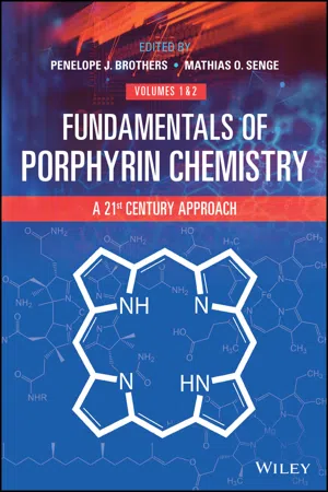 eBook - PDF
Fundamentals of Porphyrin Chemistry
A 21st Century Approach
- Penelope J. Brothers, Mathias O. Senge, Penelope J. Brothers, Mathias O. Senge(Authors)
- 2022(Publication Date)
- Wiley(Publisher)
In fact, there are no other known metalloproteins that can be probed with so many distinct spectroscopic tools. As a result, the literature on heme enzymes is substantially larger than any other metalloenzymes and can be overwhelming. The goal of this chapter is to summarize the structure–function correlations discovered for a few important heme enzymes to serve as exemplars. 16.2 Myoglobin and Hemoglobin 16.2.1 Function and Structure Oxygen has poor solubility in aqueous media (40 mg L −1 at 25 ∘ C) and therefore cannot be carried to tissues in sufficient quantity if it is simply dissolved in blood serum. The diffusion of oxygen through tissues is also insufficient to serve the cellular demand. The evolution of complex multicellular organisms led to the evolution of proteins that could transport and store oxygen. Hemoglobin (Hb, molecular weight 64 500 g mol −1 = 64.5 kDa) is the protein that carries oxygen from the lungs through the arteries to the tissues, and on the return trip, 712 16 Heme Proteins – Structure and Function takes the carbon dioxide that has been produced in the cells, as a result of cellular respira- tion, through the veins back to the lungs. Hemoglobin is found in the red blood cells. Each red blood cell contains about 280 million Hb units, comprising approximately one third of the mass of a mammalian red blood cell. Myoglobin (Mb, 16.7 kDa) is a relatively simple oxygen-binding protein found in almost all mammals, primarily in muscle tissue. In the late 1950s, the first X-ray crystal structures of Mb and Hb were solved, and currently crystal struc- tures of Mb are available in several ligation states and conformations. Hb is a tetrameric protein and consists of four polypeptide chains (Figure 16.3a), two alpha globin subunits, (α1 and α2 chains), and two beta globin subunits, or (β1 and β2 chains), while Mb is a monomeric protein (Figure 16.3b).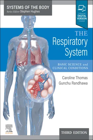 eBook - ePub
The Respiratory System E-Book
Basic science and clinical conditions
- Caroline R Thomas, Gunchu Randhawa, Stephen H. Hughes(Authors)
- 2022(Publication Date)
- Elsevier(Publisher)
2 /L blood.Haemoglobin
Hb has remarkable O2 -carrying properties which are related to its molecular structure (Fig. 8.1 ). Each Hb molecule consists of a protein (globin) and haem (protoporphyrin and ferrous iron). The globin is made up of four polypeptide chains, each carrying a haem group, which means there are four sites, each capable of carrying one O2on each Hb molecule. It is conceptually useful to consider each Hb molecule as having four ‘hooks’. On each hook can hang one O2 . This structure explains many of the properties of Hb, as we will see below.Each of the four polypeptide chains can vary in a way which will alter the O2 -carrying properties of the blood. The chains are named according to their structure. Adult Hb consists of two α and two β chains, with 141 and 146 amino acid residues per chain, respectively. Thus each Hb molecule has 574 amino acids and four haems, which gives the molecule a weight of about 64 500 Da. The various other forms of normal and abnormal Hb comprise other combinations of polypeptide chain structures. These are discussed under ‘Haemoglobin variants’, below. Men have about 150 g L− 1Hb in their blood, women about 130 g L− 1.Oxygen combination with Haemoglobin
This reversible reaction can be summarised as follows:which will be driven to the right (to oxyHaemoglobin, HbO2 ) by increased PO2 and to the left (to deoxyHaemoglobin, Hb) by low PO2 .Fig. 8.1The structure of Haemoglobin. Each of the four globin chains (two α chains and two β chains) is made up of a spiral of just over 100 amino acids. Each chain is attached to an iron (Fe2+ )- containing haem group.Each haem group can carry a molecule of oxygen (O2 ), so each Haemoglobin molecule has four ‘hooks’, each of which can carry one O2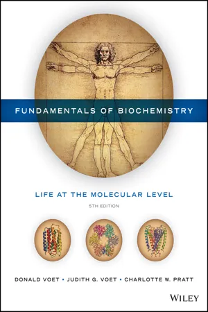 eBook - PDF
Fundamentals of Biochemistry
Life at the Molecular Level
- Donald Voet, Judith G. Voet, Charlotte W. Pratt(Authors)
- 2016(Publication Date)
- Wiley(Publisher)
Animals that are too large (>1 mm thick) for simple diffusion to deliver suffi- cient oxygen to their tissues have circulatory systems containing hemoglobin or a protein of similar function that does so (Box 7-1). In vertebrates, hemoglo- bin is contained in erythrocytes (red blood cells; Greek: erythros, red + kytos, a hollow vessel), 6–9 μm diameter, biconcave disk-shaped cells that contain ∼34% hemoglobin by weight. In mammals, they are devoid of intracellular organelles (e.g., nuclei and mitochondria), thus providing more space for hemoglobin. Mammalian hemoglobin, as we saw in Fig. 6-33, is an α 2 β 2 tetramer (a dimer of αβ protomers). The α and β subunits are structurally and evolutionarily related to each other and to myoglobin. The structure of hemoglobin was deter- mined by Max Perutz (Box 7-2). Only about 18% of the residues are identical in myoglobin and in the α and β subunits of hemoglobin, but the three polypep- tides have remarkably similar tertiary structures (hemoglobin subunits follow the myoglobin helix-labeling system, although the α chain has no D helix). The αβ protomers of hemoglobin are symmetrically related by a 2-fold rotation (i.e., a rotation of 180° brings the protomers into coincidence). In addition, hemoglobin’s structurally similar α and β subunits are related by an approximate 2-fold rotation (pseudosymmetry) whose axis is perpendicular to that of the exact 2-fold rotation. Thus, hemoglobin has exact C 2 symmetry and pseudo-D 2 186 Max Perutz (1914–2002) The determination of the three-dimensional structures of proteins has become so commonplace that it is difficult to appreciate the challenges that faced the first protein crystallographers. Max Perutz was a pioneer in this area, spending many years determining the structure of hemoglobin at atomic resolution and then using this information to explain the physi- ological function of the protein. In 1934, two years before Perutz began his doctoral studies in Cambridge, J.D. eBook - ePub
Sports Training Principles
An Introduction to Sports Science
- Dr. Frank W. Dick O.B.E.(Author)
- 2014(Publication Date)
- Bloomsbury Sport(Publisher)
3 . The red colouring is due to its Haemoglobin content, which is a combination of a protein (globin) and a red pigment (haematin). Muscle Haemoglobin is called myoglobin. The red pigment contains iron, which readily combines with oxygen. This combination is a very loose affair and the oxygen can be just as easily ‘disconnected’ or cast free. Herein lies the oxygen transporting property of blood and the obvious importance of dietary iron. However, excessive iron will not increase the oxygen-carrying capacity of the blood. Iron absorption is tightly controlled by the body’s requirements. When these are met, absorption through the intestine wall ceases and the excess iron is expelled in the faeces.In men, the average Haemoglobin (Hb) content is 15.8g/100ml blood, while in women it is 13.9g/100ml blood. To be more precise, normal values may be found within the range 14–18g/100ml blood for men and 11.5–16g/100ml for women. As 1g of fully saturated Haemoglobin combines with 1.34ml oxygen, so Haemoglobin may be used as an index of the oxygen-carrying capacity of the blood. Occasionally Haemoglobin content is expressed as a percentage, but this can be a little confusing since 100 per cent may be normal for one investigator but not for another. Moreover, there appears to be different ‘normal’ values according to age, gender, nationality, geographic location, and so on. Consequently, one must check the meaning of 100 per cent before evaluating the Haemoglobin count of an athlete.FIGURE 5.1 Summary of blood composition and functionThe idea of a normal range seems much less problematic. Information on Haemoglobin status is presented in the Edinburgh Royal Infirmary Bioprofile, as in table 5.1 eBook - PDF
- Donald Voet, Charlotte W. Pratt, Judith G. Voet(Authors)
- 2014(Publication Date)
- Wiley(Publisher)
Oxygenation alters the electronic state of the Fe(II)–heme complex, as indicated by its color change from dark purple (the color of hemo- globin in venous blood) to brilliant scarlet (the color of hemoglobin in arterial blood). Under some conditions, the Fe(II) of myoglobin or hemoglobin becomes oxidized to Fe(III) to form metmyoglobin or methemoglobin, respectively; these proteins are responsible for the brown color of old meat and dried blood. In addition to O 2 , certain other small molecules such as CO, NO, and H 2 S can bind to heme groups in proteins. These other compounds bind with much higher affinity than O 2 , which accounts for their toxicity. CO, for ex- ample, has 200-fold greater affinity for hemoglobin than does O 2 . Myoglobin Binds O 2 to Facilitate Its Diffusion. Although myoglobin was originally thought to be only an oxygen-storage protein, it is now apparent that its major physiological role is to facilitate oxygen diffusion in muscle (the most rapidly respiring tissue under conditions of high exertion). The rate at which O 2 can diffuse from the capillaries to the tissues is limited by its low solubil- ity in aqueous solution (10 4 M in blood). Myoglobin increases the effective solubility of O 2 in muscle cells, acting as a kind of molecular bucket brigade to boost the O 2 diffusion rate. The oxygen-storage function of myo- globin is probably significant only in aquatic mammals such as seals and whales, whose muscle myoglobin concentrations are around 10-fold greater than those in terrestrial mammals (which is one reason why Kendrew chose the sperm whale as a source of myoglobin for his X-ray crystallographic stud- ies). Nevertheless, mice in which the gene for myoglobin has been “knocked out” appear to be normal, although their muscles are lighter in color than those of wild-type mice.
- Paul J. Ponganis(Author)
- 2015(Publication Date)
- Cambridge University Press(Publisher)
4.1 Hemoglobin structure and function Hemoglobin (Hb) in diving birds and mammals is similar in structure to that of other mammals and birds in that it is composed of four polypeptide chains, each with an iron- containing heme group that can bind an O 2 molecule. When all four heme sites are bound with O 2 , Hb is fully saturated. At 100% saturation, there are 1.34 ml O 2 per gram of Hb. The concentration of Hb in blood is the primary determinant of blood O 2 content because the solubility of O 2 in blood at body temperature is quite low (0.00124 mM mm Hg –1 or 0.003 ml O 2 dl –1 mm Hg –1 ) (Powell, 2000, West, 1972). There are two forms of Hb which cannot bind to O 2 . Methemoglobin, which contains iron in the ferric state (Fe +++ ) as opposed to the usual ferrous state (Fe ++ ), is unable to bind O 2 , but is usually found in only minimal concentrations (Reeder, 2010). Carboxyhemoglobin is also incapable of binding O 2 , but again is usually only present in very low concentrations in most animals. It is formed when carbon monoxide, which has 240 times greater affinity for Hb than O 2, is present in the blood (West, 1972). 4.2 O 2 –hemoglobin dissociation curves The O 2 –Hb dissociation curve (Fig. 4.1) describes the reversible binding of O 2 to Hb as a function of Hb saturation and the partial pressure of O 2 (P O2 ). Its sigmoidal shape is due to allosteric interactions and cooperativity between the four polypeptide subunits of the Hb molecule (Powell, 2000, West, 1972). P 50 , the P O2 at which Hb is 50% saturated, is used as an index of Hb’s affinity for O 2 , and its reference value is usually taken at normal body temperature and blood pH (pH 7.4 in mammals and 7.5 in birds). The O 2 – Hb dissociation curve (and the P 50 ) can be shifted to the left or right by changes in three primary factors that regulate the O 2 affinity of Hb. eBook - PDF
- Anatoly Bezkorovainy, Max E. Rafelson(Authors)
- 1996(Publication Date)
- CRC Press(Publisher)
Of al l hemoglobin-type protein s known , th e simples t on e fro m a structural point o f vie w i s myoglobin . Thi s sper m whal e protei n consist s o f a singl e Proteins with Biological Activity: Blood Protein s 157 480 50 0 52 0 54 0 56 0 58 0 60 0 62 0 64 0 Wavelength (nm) Figure 7. 1 Absorptio n spectra o f various hemoglobin s i n the 480-64 0 n m range . Me t Hb, 0 2 H b , COHb, H Hb and SulfHb designates methemoglobin, oxygenated hemoglobin, carbon monoxid e hemoglobi n (carboxyhemoglobin) , deoxyhemoglobin an d sulfhemo -globin, respectively . (Reproduce d wit h permissio n fro m Mora n RF , Fallon K D . Oxygen saturation, conten t an d th e dyshemoglobins : Part I . Cli n Che m New s January:! 1, 1990. ) polypeptide chai n with 15 3 amin o acid s an d a singl e hem e molecul e fo r a tota l molecular weight o f about 18,000 . I t is quite compact , measurin g 44 x 4 4 x 2 5 A. Myoglobin i s th e mos t primitiv e hemoglobin-lik e protei n fro m a n evolutionar y point o f view . I t is a n intracellula r protein whos e functio n i s t o stor e oxyge n i n the cell s fo r metabolic purposes . The structur e of hem e i s illustrate d in Figur e 7.2 . Th e molecule consist s o f four pyrrole-lik e ring structure s (se e structur e I, Figure 7.2 ) joine d b y methen e bridges (=C-) , plu s variou s side chains : methyl, vinyl , an d propyl . It is a planar molecule. Associate d with nitroge n atom s o f th e rin g i s a ferrous iron ion (Fe 2 + ); two nitroge n atom s hav e los t thei r protons , thu s carryin g on e negativ e charg e each, an d thes e the n for m electrostati c interaction s wit h th e positivel y charge d iron. Th e other tw o nitroge n atom s for m coordinate-covalent bond s wit h iron . If iron is removed fro m heme, th e resul t is protoporphyrin IX (structure IV, Figur e 7.2). Th e chemical prototype o f al l porphyrin-type compounds i s porphin (struc-ture III , Figur e 7.2) .
Index pages curate the most relevant extracts from our library of academic textbooks. They’ve been created using an in-house natural language model (NLM), each adding context and meaning to key research topics.