Chemistry
Quaternary Structure of Protein
The quaternary structure of a protein refers to the arrangement of multiple protein subunits to form a functional protein complex. This structure is stabilized by various interactions such as hydrogen bonds, disulfide bonds, and hydrophobic interactions between the subunits. The quaternary structure is essential for the overall function and stability of many proteins.
Written by Perlego with AI-assistance
Related key terms
1 of 5
11 Key excerpts on "Quaternary Structure of Protein"
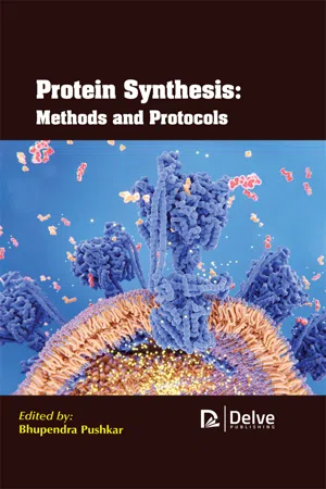 eBook - PDF
- Bhupendra Pushkar(Author)
- 2020(Publication Date)
- Delve Publishing(Publisher)
The ensemble of formations and folds in a single linear chain of amino acids is sometimes referred to as a polypeptide. And this contains the tertiary structure of a protein. Ultimately, the quaternary structure of a protein refers to those macromolecules which have multiple polypeptide chains or subunits. The shape that is finally adopted by a newly synthesized protein is typically the most energetically favorite one. As the folding of proteins takes place, they tend to test a variety of conformations before reaching their final form which is said to be unique and compact. The stabilization of the folded proteins takes place by a large number of non – covalent bonds between amino acids. Protein Synthesis: Methods and Protocols 6 Along with this, there are chemical forces also in between a protein and its immediate environment which contribute to the protein shape and stability. Let’s take an example of the proteins which are dissolved in the cell cytoplasm. These proteins have hydrophilic (water-loving) chemical groups on their surfaces whereas, on the other hand, their hydrophobic (water-averse) elements is tucked inside. Contrastingly, there are proteins that are inserted into the cell membranes and these proteins display some hydrophobic chemical groups on their surface, particularly in those regions or areas where the protein surface is exposed to membrane lipids. However, it is important to consider that the folded proteins are not frozen into shape. Instead of this, there are atoms within these proteins that have the capability to make small movements. Although the proteins are considered as macromolecules, they are too small to be visualized, even by using a microscope. Therefore,scientists are required to use some of the indirect methods to figure out what they look like and their folding process. There is a most commonly used method for studying the protein structures and that is named X-ray crystallography. eBook - PDF
- BIOTOL, B C Currell, R C E Dam-Mieras(Authors)
- 2013(Publication Date)
- Butterworth-Heinemann(Publisher)
A protein's structure will be the sum of secondary and tertiary structures, since it refers to the overall 3-dimensional shape, covering the path that the polypeptide chain follows as well as the position that the sidechains occupy. Quaternary structure This only occurs when more than one polypeptide chain associate together to form the biologically functional molecule. Each polypeptide is described as a subunit, or protomer. Subunits then associate to form the active oligomer. Proteins in which there is only a single polypeptide (such as myoglobin) thus do not have a quaternary structure. Haemoglobin, which consists of 4 polypeptides (2 of one primary sequence, called a-subunits; 2 of a different, although similar, sequence, called ß-subunits) is described as having the quaternary structure Ofcßz sub-unit, protomer Proteins 47 3.3.2 Stabilising forces in proteins We have already said that a protein adopts one particular conformation, which is biologically active, rather than numerous alternative forms (most of which would be inactive). How does this happen? The reason is that the so-called native conformation is more stable than all other possible conformations and is a consequence of a large number of individually weak non-covalent bonds or interactions. Since these are very important, not only for protein structure but also for other macromolecules (eg nucleic acids) and for the interactions between molecules (eg enzymes and substrates; hormones and receptors) we shall review them. Typical strengths of these bonds are shown in Table 3.3, together with that of the peptide bond. Note also that proteins may be stabilised by the formation of disulphide bridges, which will also be discussed. Bond Covalent (peptide, disulphide) Hydrogen (C=O .... HN-) Ionic (-COO...H + 3 N-) Hydrophobie (eg overlapping phenyl rings) Van der Waals (transient dipoles) Bond strength*, kJ.mol 1 Approx. eBook - ePub
- Leonard J. Banaszak(Author)
- 2000(Publication Date)
- Academic Press(Publisher)
CHAPTER 5 Quaternary Structure of ProteinsINTRODUCTION
Hemoglobin (Hb) was more than just the first protein whose structure was determined by crystallography—it was the first oligomeric protein. So, in addition to the information that described the conformation of the α and β chains, the arrangement of subunits in a single Hb tetramer was also determined. From these early results and those of other researchers working on spherical viruses, a set of rules governing the assembly of subunits into limited aggregates evolved. The rules center around symmetry arguments, and make it possible to predict, or at least set some usable principles to describe, the quaternary structure of any one oligomeric protein.Such principles form the basis of any discussion of quaternary structure. They are described as follows:1. The interactions that govern the association of subunits into higher aggregates are the same as those that cause proteins to fold. Entropic factors are important, as are all of the noncovalent bonding forces.2. In oligomeric globular proteins, these thermodynamic driving forces result in the association of subunits in a constant stereochemical sense to form one unique molecule. The phrase constant stereochemical sense derives from specific noncovalent bonds that are formed to minimize the free energy of the system. This implies a form of molecular recognition in the same sense that a substrate recognizes the active site of an enzyme.3. Association of polypeptide chains takes two forms: (a) closed association to form an oligomer and (b) continuous association to form a polymer, for example, F-actin.ASSOCIATION OF PROTEIN SUBUNITS
Figure 5.1 shows three examples of a protein protomer aggregating to form a hypothetical oligomer. The three forms are different because of the types of contacts that are formed. Each type of contact represents a different set of noncovalent bonds between the subunits. Note that in Fig. 5.1A , an oligomer forms with contacts between points u and w on each protomer. Association continues with the u–w contact beyond the dimer level; however, this form of association stops with the formation of a trimer, because steric limitations prevent the addition of a fourth subunit with u–w No longer available |Learn more
- (Author)
- 2014(Publication Date)
- College Publishing House(Publisher)
The shape into which a protein naturally folds is known as its native conformation. Although many proteins can fold unassisted, simply through the chemical properties of their amino acids, others require the aid of molecular ______________________________ WORLD TECHNOLOGIES ______________________________ chaperones to fold into their native states. Biochemists often refer to four distinct aspects of a protein's structure: • Primary structure : the amino acid sequence. • Secondary structure : regularly repeating local structures stabilized by hydrogen bonds. The most common examples are the alpha helix, beta sheet and turns. Because secondary structures are local, many regions of different secondary structure can be present in the same protein molecule. • Tertiary structure : the overall shape of a single protein molecule; the spatial relationship of the secondary structures to one another. Tertiary structure is generally stabilized by nonlocal interactions, most commonly the formation of a hydrophobic core, but also through salt bridges, hydrogen bonds, disulfide bonds, and even post-translational modifications. The term tertiary structure is often used as synonymous with the term fold . The tertiary structure is what controls the basic function of the protein. • Quaternary structure : the structure formed by several protein molecules (polypeptide chains), usually called protein subunits in this context, which function as a single protein complex. Proteins are not entirely rigid molecules. In addition to these levels of structure, proteins may shift between several related structures while they perform their functions. In the context of these functional rearrangements, these tertiary or quaternary structures are usually referred to as conformations, and transitions between them are called conformational changes. eBook - PDF
- Ulo Langel, Benjamin F. Cravatt, Astrid Graslund, N.G.H. von Heijne, Matjaz Zorko, Tiit Land, Sherry Niessen(Authors)
- 2009(Publication Date)
- CRC Press(Publisher)
35 3 Structural Organization of Proteins Matjaž Zorko In 1951, while lecturing at Stanford University, the Danish biochemist Kaj Ulrik Linderstrøm-Lang was the first to propose the four-level, three-dimensional orga-nization of protein structures. These structures can be described as follows: 1. The primary structure: the covalent chemical structure of the polypeptide chain or chains in a given protein, that is, the number and sequence of the amino acid residues linked together by the peptide bonds. 2. The secondary structure: any such folding that is brought about by linking the carbonyl and the imides groups of the backbone together by means of hydro-gen bonds. This structure includes helices, sheets, turns, loops, and so on. 3. The tertiary structure: the organization of the secondary structures linked by “looser segments” of the polypeptide chain stabilized primarily by the side-chain interactions. The disulfide bonds are included in this level. 4. The quaternary structure: the aggregation of the separate polypeptide chains into a functional protein. CONTENTS 3.1 The Primary Structure ..................................................................................... 36 3.2 The Secondary Structure ................................................................................. 37 3.2.1 The Helices ......................................................................................... 39 3.2.1.1 The α -Helix ......................................................................... 40 3.2.1.2 The Other Helices ................................................................ 44 3.2.2 The β -Structure ................................................................................... 45 3.2.3 The Turns ............................................................................................ 47 3.2.4 Poly-Pro and Poly-Gly ........................................................................
- Frank H. Stillinger(Author)
- 2015(Publication Date)
- Princeton University Press(Publisher)
Case ( β 3) presents another example, whose tetrameric quaternary geometry consists of two identical dimers, each one of which in-cludes hydrogen bonding between side-by-side beta sheets connecting separate protein monomers (a situation that is known as a “cross-β conformation”). A third example is the bacterial enzyme phosphofructokinase (from Bacillus stearothermophilus ), consisting of four identical protein units, each containing both alpha-helical and beta-sheet secondary structures [Evans et al., 1981]. In addition to these quaternary combinations of a small number of individual protein mole-cules, much larger protein aggregates of indefinite size also play vital roles in living organisms. Structural proteins such as actin [Otterbein et al., 2001], the collagens [Di Lullo et al., 2002], keratins [Kreplak et al., 2004], tubulins [Nogales et al., 1998], and silk fibroins [Sashina et al., 2006] are proteins that fall into this category. Although structural details vary, the individual molecules in these substances tend to be organized to constitute extended fibers rather than glob-ular tertiary structures, a basic feature that contributes to the necessary mechanical properties demanded by their biological roles. Protein Folding Phenomena 451 Unfortunately, there are also pathological cases of quaternary aggregation of large numbers of “misfolded” protein molecules [Dobson, 2003; Selkoe, 2003; Chiti and Dobson, 2006]. This situation underlies the family of “amyloid diseases,” also known as “amyloidoses.” Besides “mad cow” disease, this category includes several serious human disorders such as Alzheimer’s disease, Parkinson’s disease, Creutzfeldt-Jakob disease, Huntington’s disease, and Type II diabetes. The anatomical reasons for occurrence of these pathologies, and how they might best be treated and prevented, remain important research objectives. eBook - PDF
- Clarence H. Suelter(Author)
- 2009(Publication Date)
- Wiley-Interscience(Publisher)
We have not attempted a comprehensive review of the many publications describing work in which monoclonal antibodies and limited proteolysis have played a major part, but have tended to focus on more recent applications. The interested reader is referred to a previous publication (2 1 ) that provides additional discussion on applications of monoclonal antibodies to structure-function studies. 1. UNDERLYING CONCEPTS 2.1. Protein Structure A. QUATERNARY THE STRUCTURAL HIERARCHY: PRIMARY, SECONDARY, TERTIARY, One frequently used approach (22) has been to view protein structure as a progression from an essentially linear representation (the primary structure, defined as the amino acid sequence) through localized hydrogen bonded structures such as a helices, f3 pleated sheets, or reverse turns (secondary structures) which in turn become associated (folded) to yield a specific three- dimensional array, referred to as the tertiary structure. In some proteins, two or more such folded subunits may associate to give the final oligomeric (quaternary) structure in which biological activity is fully expressed. Such distinctions can be useful for discussion purposes, but should not be taken to imply true independence. Since the classic work by Anfinsen and his colleagues (23), it has been recognized that the amino acid sequence encodes the information necessary for development of higher orders of structure, and this provides a basis for attempts to predict secondary structure from amino acid sequence (e.g., 24). Though presently available methods for secondary structural prediction do not boast a remarkable accuracy (25, 26), they can certainly prove useful, particularly when several methods are used in combination (26). Further improvement in predictive accuracy can be expected as relationships between amino acid sequence and specific secondary structural features are more fully understood (27, 28). eBook - ePub
- David P. Clark(Author)
- 2009(Publication Date)
- Academic Cell(Publisher)
When the signal molecule is bound, it changes the shape of its own domain (Fig. 7.16). The change in conformation is then transmitted to the DNA-binding domain, which also changes shape. Thus, although they fold separately, domains do interact physically. Figure 7.16 Interactions between Protein Domains to Activate a DNA-Binding Site A) The unfolded protein shows two domains (I and II) and a linker region. B) When folded, both domain I and domain II form binding sites. C) A signal molecule binds to domain I and changes its conformation. The interaction between the two domains triggers domain II to change shape so opening up its binding site. Quaternary Structure of Proteins Many proteins consist of several individual polypeptide chains. This is especially true of proteins whose total molecular weight is much greater than 50,000 daltons (i.e. around 400 amino acids). [Although occasional polypeptide chains are found with a 1,000 or more amino acids, they are relatively rare.] The assembly of these multiple subunits yields the quaternary structure (Fig. 7.17). (Proteins with only one polypeptide chain have no quaternary structure.) The subunits, or protomers, are usually present as an even number, most often two or four. The terms dimer, trimer, tetramer, oligomer and multimer refer to structures with two, three, four, few/several and multiple subunits, respectively. Less than 10 percent of multimeric proteins have an odd number of subunits. The subunits may be all identical or all different or several each of two (or more) different types. For example, the lactose repressor consists of four identical subunits, whereas hemoglobin has two α-subunits and two β-subunits. The prefixes homo- (same) and hetero- (different) are sometimes used to indicate whether the subunits are the same or different eBook - PDF
Mobility and recognition in cell biology
Proceedings of a FEBS Lecture Course held at the University of Konstanz, West Germany, September 6–10, 1982
- Horst Sund, Cees Veeger, Federation of European Biochemical Societies, Horst Sund, Cees Veeger, Federation of European Biochemical Societies(Authors)
- 2019(Publication Date)
- De Gruyter(Publisher)
SELF-ORGANIZATION OF OLIGOMERIC PROTEINS Folding and Recognition of Polypeptide Chains upon Quaternary Structure Formation Rainer Jaenicke Institut für Biophysik und Physikalische Biochemie Universität Regensburg D-8400 Regensburg, Germany Introduction The autonomous transition from the one-dimensional amino acid sequence to the three-dimensional spatial organization of pro-tein molecules is called folding (1). Association gener-ating the next higher level in the hierarchy of structures re-quires mobility and recognition of the subunits involved. Folding and- association must be properly coordinated because the formation of the native quaternary structure requires the surface of the structured monomers to be preformed in the correct way such that specificity is achieved. The limits of specificity, i.e. the occurrence of hybrid or chimeric assem-blies, reflect the flexibility of the tertiary structure of a given sample of polypeptide chains. The code by which the amino acid sequence of a protein spec-ifies its folding is unknown so far. Due to the vectorial character of the translation process kinetic constraints are involved in the acquisition of the three-dimensional structure. The experimentally observed kinetics of folding clearly contra-dict a purely thermodynamic concept: Comparing the time scale of in vivo folding to the time required to probe all confor-mations in search of the global energy minimum, a difference of ^60 orders of magnitude is predicted. Nucleation, exclu-ded volume effects, domain folding etc may be considered to explain the discrepancy (2). Mobility and Recognition in Cell Biology © 1 9 8 3 by Walter de Gruyter & Co., Berlin • N e w York 68 R. Jaenicke Folding in vivo - Refolding in vitro The pathway of folding should reveal the folding mechanism. Un-fortunately at present no direct approach is available to study the acquisition of the native structure of the nascent polypep-tide chain either in vivo or in vitro. eBook - PDF
- Hans Neurath(Author)
- 2012(Publication Date)
- Academic Press(Publisher)
Most of the proteins listed in Table II are composed of identical sub-TABLE Distribution of Subunit Stoichiometries Number of Number of proteins with desig- Number of proteins with designated subunits nated number of identical subunits« number of nonidentical subunits 2 3 4 5 6 7 8 9 10 11 12 96 7 77 0 26 0 11 0 2 0 7 13 3 17 0 1 0 2 0 1 0 3 ° Summed from entries in Table II. 5. Quaternary Structure of Proteins 337 TABLE IV Constitution of Proteins Composed of Nonidentical Subunits Subunit Protein Molecular weight composition Rat pituitary leutinizing hormone Neurospora malate dehydrogenase Lactose synthetase Aldose reductase Hemoglobin T u -T 8 Complex Bacterial luciferase Troponin Bovine procarboxypeptidase A Serum high density lipoprotein Succinate dehydrogenase Tuberlin Aspartokinase Pasteurella protein toxin B Fructose diphosphatase Protein kinase Succinyl-CoA synthetase Serratia anthranilate synthetase Tryptophan synthetase Molybdoferredoxin E. colt carbamyl phosphate synthetase Histidine decarboxylase Dipeptidyl transferase E. No longer available |Learn more
- Dagmar Klostermeier, Markus G. Rudolph(Authors)
- 2018(Publication Date)
- CRC Press(Publisher)
Such domains may not necessarily functionally interact, but the domain movements enable large conformational changes of the protein, which is important for function. Titin is an extreme example of a multi-domain protein where the domains apparently do not functionally cross-talk. This vertebrate muscle-organizing protein has the largest single poly-peptide chain in humans with 27 000–38 000 residues depending on the titin isoform. Among oth-ers, titin harbors a kinase domain, 152 Ig domains, and 132 fibronectin-like domains ( β -sandwiches similar to Ig domains). While it is often observed that individual domains can be isolated from multi-domain proteins and are stable in solution, for many other proteins the linker is important for domain stabilization. 16.2.5 Q UATERNARY S TRUCTURE & P ROTEIN -P ROTEIN I NTERACTIONS Several polypeptide chains that assemble into higher-order oligomers form a quaternary structure. A weak correlation exists between the molecular mass of a protein and its tendency to oligomerize: monomeric proteins generally have rather small molecular masses of around 30 kDa, whereas larger proteins > 50 kDa molecular mass tend to form oligomers. Assembly into a quaternary structure is a general strategy to increase protein functionality and stability, or to enable protein regulation. Oligomers frequently undergo cycles of large conformational changes that may not be possible for monomers. Oligomerization can be made irreversible by covalent crosslinks such as disulfide bonds, which results in very stable assemblies. If the assembly is reversible, the oligomer is normally in dynamic equilibrium with its constituent monomers. A survey of crystal structures of oligomeric proteins ( Table 16.7 ) has shown that homo-oligomers are more frequent than hetero-oligomers, small stoichiometries are preferred over large stoichiometries, and even-numbered components are preferred over odd-numbered components.
Index pages curate the most relevant extracts from our library of academic textbooks. They’ve been created using an in-house natural language model (NLM), each adding context and meaning to key research topics.