Chemistry
Tertiary Structure of Protein
The tertiary structure of a protein refers to the three-dimensional arrangement of its secondary structural elements, such as alpha helices and beta sheets, as well as any loops and folds. This structure is stabilized by various interactions, including hydrogen bonds, disulfide bonds, hydrophobic interactions, and electrostatic forces. The tertiary structure is crucial for the protein's function and stability.
Written by Perlego with AI-assistance
Related key terms
1 of 5
12 Key excerpts on "Tertiary Structure of Protein"
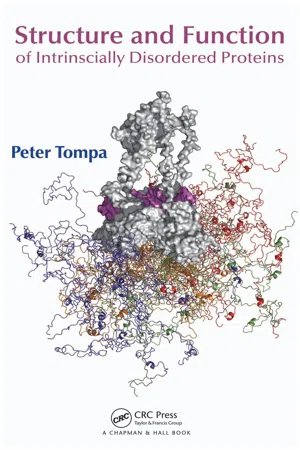
- Peter Tompa, Alan Fersht(Authors)
- 2009(Publication Date)
- Chapman and Hall/CRC(Publisher)
This situation may be treated by applying a more thorough set of definitions, as suggested in the dictionary of protein secondary struc-ture (DSSP) approach (Kabsch and Sander 1983). The DSSP scheme is founded not on angles but on the presence/absence of H-bonds, defined by a threshold value –0.5 kcal/mole of interaction calculated from partial charges and interatomic distances. Two elementary H-bond types are defined, and a turn occurs when there is a H-bond between C=O of residue (i) and NH of residue (i + n), where n = 3, 4, or 5, whereas a bridge is defined between two (parallal or antiparallel) stretches of tripeptides if the actual residues (i and j) that form a H-bond are more remote in sequence than in the case of the turn. A minimal helix is then defined as two consecutive n-turns, whereas a longer helix is described as overlapping minimal helices (an α -helix, for example, is described as repeating 4-turns). A ladder is defined as a set of consecu-tive bridges of identical type, whereas a sheet is defined as one or more ladders con-nected by shared residues. The extraction of such patterns from structures is easily automated. 1.5.2 Tertiary Structure Strictly speaking, the tertiary structure of a protein is the folding of its polypeptide chain in 3-D space, described by the coordinates of all its atoms. Because this descrip-tion is difficult to interpret, usually a simplified description of the topology of its sec-ondary structural elements is used instead. In the case of certain elongated fibrous proteins, the structure is built up of a simple secondary structural element. The long fiber of α -keratin, for example, is composed of 10 Structure and Function of Intrinsically Disordered Proteins a structural unit of two long α -helices twisted around each other (termed a two-stranded coiled coil). Fibroin and β -keratin found in silk fibers are composed of stacked antipar-allel β -sheets. eBook - PDF
- Bhupendra Pushkar(Author)
- 2020(Publication Date)
- Delve Publishing(Publisher)
The ensemble of formations and folds in a single linear chain of amino acids is sometimes referred to as a polypeptide. And this contains the tertiary structure of a protein. Ultimately, the quaternary structure of a protein refers to those macromolecules which have multiple polypeptide chains or subunits. The shape that is finally adopted by a newly synthesized protein is typically the most energetically favorite one. As the folding of proteins takes place, they tend to test a variety of conformations before reaching their final form which is said to be unique and compact. The stabilization of the folded proteins takes place by a large number of non – covalent bonds between amino acids. Protein Synthesis: Methods and Protocols 6 Along with this, there are chemical forces also in between a protein and its immediate environment which contribute to the protein shape and stability. Let’s take an example of the proteins which are dissolved in the cell cytoplasm. These proteins have hydrophilic (water-loving) chemical groups on their surfaces whereas, on the other hand, their hydrophobic (water-averse) elements is tucked inside. Contrastingly, there are proteins that are inserted into the cell membranes and these proteins display some hydrophobic chemical groups on their surface, particularly in those regions or areas where the protein surface is exposed to membrane lipids. However, it is important to consider that the folded proteins are not frozen into shape. Instead of this, there are atoms within these proteins that have the capability to make small movements. Although the proteins are considered as macromolecules, they are too small to be visualized, even by using a microscope. Therefore,scientists are required to use some of the indirect methods to figure out what they look like and their folding process. There is a most commonly used method for studying the protein structures and that is named X-ray crystallography. eBook - PDF
- Ulo Langel, Benjamin F. Cravatt, Astrid Graslund, N.G.H. von Heijne, Matjaz Zorko, Tiit Land, Sherry Niessen(Authors)
- 2009(Publication Date)
- CRC Press(Publisher)
35 3 Structural Organization of Proteins Matjaž Zorko In 1951, while lecturing at Stanford University, the Danish biochemist Kaj Ulrik Linderstrøm-Lang was the first to propose the four-level, three-dimensional orga-nization of protein structures. These structures can be described as follows: 1. The primary structure: the covalent chemical structure of the polypeptide chain or chains in a given protein, that is, the number and sequence of the amino acid residues linked together by the peptide bonds. 2. The secondary structure: any such folding that is brought about by linking the carbonyl and the imides groups of the backbone together by means of hydro-gen bonds. This structure includes helices, sheets, turns, loops, and so on. 3. The tertiary structure: the organization of the secondary structures linked by “looser segments” of the polypeptide chain stabilized primarily by the side-chain interactions. The disulfide bonds are included in this level. 4. The quaternary structure: the aggregation of the separate polypeptide chains into a functional protein. CONTENTS 3.1 The Primary Structure ..................................................................................... 36 3.2 The Secondary Structure ................................................................................. 37 3.2.1 The Helices ......................................................................................... 39 3.2.1.1 The α -Helix ......................................................................... 40 3.2.1.2 The Other Helices ................................................................ 44 3.2.2 The β -Structure ................................................................................... 45 3.2.3 The Turns ............................................................................................ 47 3.2.4 Poly-Pro and Poly-Gly ........................................................................ eBook - PDF
Biochemistry
An Integrative Approach
- John T. Tansey(Author)
- 2019(Publication Date)
- Wiley(Publisher)
• Secondary structures are stabilized by hydrogen bonds in the peptide backbone, and can be formed by many different amino acids. • Tertiary structures describe the interaction of different elements of helix and sheet to form a complex structure. • Tertiary structure represents the complete folding of a single polypeptide into a functional protein. • Examples of tertiary motifs include the four-helix bundle and the Greek key motif. • Tertiary structures are stabilized by a combination of many weak forces; these include hydrogen bond- ing, dipole–dipole interactions, salt bridges, disulfide bonds, cation–π interactions, and a phenomenon known as the hydrophobic effect. • Many proteins exhibit quaternary structure interactions between and among different polypeptide chains. WORKED PROBLEM 3.3 Protein structure and disease Sickle cell anemia is a common genetic disorder. The causal mutation in sickle cell anemia is E6V, that is, the substitution of a glutamic acid for valine in the sixth position of the β chain of hemoglobin, a tetrameric protein with the stoichiometry a2β2. In the deoxygenated state, the mutant hemoglobin can polymerize, causing erythrocytes (red blood cells) to form a sickled shape and lyse, leading to the symptoms of this disease. How might this mutation affect all four levels of protein structure? Strategy Examine the information we have been given in the question and think about the four levels of protein structure. How could this alteration in amino acid sequence lead to the observed phenotype and disease? Solution Hemoglobin is a tetrameric protein. The mutation has altered the primary sequence of the β chains of hemoglobin by replacing a negatively charged glutamic acid with a hydrophobic valine residue. It is not apparent from the information provided in the question how this might affect the secondary and tertiary structure of the protein.
- Zdzislaw E. Sikorski(Author)
- 2001(Publication Date)
- CRC Press(Publisher)
The rela-tive spatial arrangement of amino acid residues is not taken into account at this level. The locations of any existing disulfide bridges should also be included here to complete a description of all covalent bonds, but in such a case, the IUPAC recommends a term “covalent structure.” The polypeptide chain is not free to take up any three-dimensional structure. Steric constraints and weak in-teractions, especially the formation of hydrogen bonds, determine that some spatial arrangements, i.e., conformations, are more stable than others. Secondary structure refers to regular recurring local conformations of adjacent amino acids in a polypeptide chain. There are a few common types of secondary structures, the most prominent being the a-helix and (3-conformation. Tertiary structure is the overall three-dimensional arrangement of the entire polypeptide chain. Usu-ally a few different types of secondary structures can be found within the ter-tiary structure of a large protein. Proteins with several polypeptide chains have one more level of structural organization: quaternary structure, which refers to the spatial relationship of the polypeptides, or subunits, within the protein. Continued advances in the understanding of protein structure and folding al-lowed two additional structural levels to be defined, both fall between sec-ondary and tertiary structural levels. A stable clustering of several elements of a secondary structure is called a supersecondary structure. These especially stable arrangements occur in many different proteins. A somewhat higher level of structural organization is the domain. This refers to a compact region that is a distinct structural and sometimes functional unit within a large polypep-tide chain. A monomeric protein may contain several domains that are often readily distinguishable within the overall structure. 3.4.2. THE PRIMARY AND SECONDARY STRUCTURES Proteins are composed of amino acids joined by amide (peptide) bonds. eBook - PDF
- Lizabeth A. Allison(Author)
- 2021(Publication Date)
- Wiley-Blackwell(Publisher)
. . . . . O O Figure 4.9 Four levels of protein structure. The primary protein structure is the sequence of a chain of amino acids. Secondary structures such as the α -helix and the β -pleated sheet are stabilized by hydrogen bonding between nearby amino acids in the chain. The secondary structure folds into a three-dimensional tertiary structure through noncovalent and covalent interactions. The quaternary structure is a protein consisting of more than one amino acid chain. 4.3 The three-dimensional structure of proteins 91 the first enzyme ever to have its structure solved by X-ray diffraction. Lysozyme is a widespread enzyme found in animal secretions such as tears and in egg white. It catalyzes the breaking of glycosidic bonds between certain residues in components of bacterial cell walls, resulting in lysis of the bacteria. Because of its catalytic properties, lysozyme is C SH HS C C H 2 H 2 H 2 H 2 S S C Figure 4.10 Disulfide bonds in tertiary folding. The backbone structure of α -chymotrypsin, an enzyme involved in digesting proteins in the small intestine, is shown (Protein Data Bank, PDB: 5CHA). Its structure contains five disulfide bonds (red bars). Cysteines are shown in light orange. The inset shows two cysteine side chains on the opposite side of a loop domain. The two thiol groups can undergo a reaction involving the loss of two hydrogens and the formation of a covalent disulfide bond between them. Chymotrypsin is activated by cleavage of the inactive precursor chymotrypsinogen, which is secreted by the pancreas. The three segments of polypeptide chain (green, light blue, and dark blue) produced by proteolytic processing remain linked by disulfide bonds. Source: Based on Protein Data Bank, PDB: 5CHA. (a) Active site (b) (c) Figure 4.11 The structure of the globular protein lysozyme. (a) A ribbon model depicts how the α -helices (coiled ribbons) and β -pleated sheets (flat arrows) present in lysozyme interact to form a globular shape. eBook - PDF
- Bhupendra Pushkar(Author)
- 2019(Publication Date)
- Delve Publishing(Publisher)
Analysis of Protein Structure 3 CONTENTS 3.1. Introduction ...................................................................................... 38 3.2. Crystallography ................................................................................. 44 3.3. Modeling .......................................................................................... 51 Protein Bioinformatics 38 3.1. INTRODUCTION To comprehend the fundamental standards of protein three-dimensional structure and the capability of their utilization in different regions of research, scholastic, or modern like pharmaceutical or biotech businesses. We first need to take a gander at the four degrees of protein structure. The distinctive secondary levels rely upon one another, together making an amazingly unpredictable system of communications somewhere in the range of thousands of iotas. The primary level is the amino acid sequence alignment – there are 20 diverse amino acids most usually found in proteins. The succession of these amino acids in a polypeptide chain basically decides the kinds of secondary structure components present, which is the second degree of association and the route by which they are masterminded in space, making basic themes and rows, which is the third degree of association. A free collapsing unit of the three-dimensional protein structure is known as a space. It is autonomous in light of the fact that spaces may frequently be cloned, communicated and refined freely of the remainder of the protein, and they may even show action, if there is any realized movement related with them. A few proteins contain one single space while others may contain a few areas. A protein space is appointed a particular kind of raw. Areas with a similar overlay could possibly be identified with one another practically. This is essentially on the grounds that Nature has re-utilized a similar overlap commonly in various settings. eBook - PDF
- BIOTOL, B C Currell, R C E Dam-Mieras(Authors)
- 2013(Publication Date)
- Butterworth-Heinemann(Publisher)
The secondary structure(s) are then linked by sections of random coil of irregular structure. extended Other helices can, in principle, occur. Think of either a more extended helix ie greater helices translation along the helix axis per residue, or a more squashed one. Short segments of a more extended helix (the 3.10 helix) are sometimes seen. The fibrous protein of connective tissue, collagen, forms an extended left-handed helix; three of these strands are combined, rope-like, to form a 'super-helix' which is stabilised by inter-chain hydrogen bonds. Proteins 59 Protein a- Keratin Myoglobin Lysozyme Carboxypeptidase Cytochromec % a- helix* 100 77 45 35 15 Table 3.4 Percentage a- helical content of some proteins. *The percentage is the proportion of the total number of amino acids which are found within a- helices. many fibrous proteins have repeated amino acid sequences It is notable that some of the fibrous (ie structural) proteins have primary structures which are 'repeats' of a simple sequence. Thus silk largely consists of a 6-residue repeat: gly-ser-gly-ala-gly-ala, whilst collagen has large amounts of glycine and proline (or hydroxyproline, which is a hydroxylated form of proline). Globular proteins, which constitute the enzymes and proteins which interact with other compounds in cells, have much more varied amino acid sequences. As a consequence, their structures are essentially infinitely varied. myoglobin structure elucidated orientation of polar and hydrophobic sidechains in proteins 3.5 Tertiary Structure of Proteins The tertiary structure describes the overall shape of the protein. This overall conformation is the consequence of the presence of any secondary structures and how they and the remainder of the chain are positioned. Each protein adopts a unique tertiary structure. This conformation will be the result of the various weak bonds which can arise. It also depends on interaction with the surrounding solvent (usually water). eBook - PDF
- S Bresler(Author)
- 2012(Publication Date)
- Academic Press(Publisher)
These results confirm the postulate that protein secondary and tertiary structures are completely determined by their primary structure, even for very complex proteins. y-Globulin, for instance, consists of four individual polypeptide chains, yet it is almost completely reconstituted by the techniques described above. Twenty years ago, Bresler and Talmud formulated a hypothesis stating that the secondary and tertiary structures of globular proteins are the result of an equilibrium among three types of molecular forces : hydrogen bonds between the peptide groups of each chain which cause the formation of helix; van der Waals forces between hydro-phobic amino acid side groups; and electrostatic forces of repulsion between charges on the surface of the molecule (72). Although the precise configuration of protein secondary and tertiary structures were unknown at that time, the ideas underlying this theory have been wholly confirmed over the intervening years. Nonetheless, estimates of the energies involved in these three types of interaction, originally made in 1949, have had to be corrected on the basis of more recent data. The energy of the hydrogen bond in a number of proteins, for example, has been found to equal 1400 cal/mole of residues participating in the helix (27). In one mole of a hypothetical protein with a molecular weight of 17,000, 60% of whose approximately 150 residues reside in helical segments, the energy of intramolecular hydrogen bonds amounts to E x = 1400 χ 0.6 χ 150 = 130,000 cal. The most important new principle introduced by the Bresler-Talmud model was that van der Waals interactions among side groups were considered to be responsible for the folding of the polypeptide chain into a compact globule. eBook - PDF
Protein Physics
A Course of Lectures
- Alexei V. Finkelstein(Author)
- 2002(Publication Date)
- Academic Press(Publisher)
Part III SECONDARY STRUCTURES OF POLYPEPTIDE CHAINS This Page Intentionally Left Blank LECTURE 7 Having dealt with elementary interactions, in this lecture we will consider the secondary structure of proteins. First of all we will discuss regular secondary structures, that is, α -helices and β -structures. These secondary structures are distinguished by regular arrangements of the main chain with side chains of a variety of conformations. The tertiary structure of a protein is determined by the arrangement of these structures in the globule (Fig. 7.1). We shall consider helices first. They can be right-handed or left-handed (Fig. 7.2) and have different periods and pitches. Right-handed (R) helices come closer to the viewer as they move counterclockwise (which corresponds to positive angle counting in trigonometry), while left-handed (L) helices approach the viewer as they move clockwise. In the polypeptide chain, major helices are stabilized by hydrogen bonds. The bonds are formed between C==O and H –– N groups of the polypeptide backbone, Figure 7.1. The secondary structures of a polypeptide chain ( α -helix and a strand of β -sheet) and the tertiary structure of a protein globule. Usually, taken together, α -and β -structures make up about a half of the chain in a globular protein. 75 76 SECONDARY STRUCTURES OF POLYPEPTIDE CHAINS R L Figure 7.2. Right-handed (R) and left-handed (L) helices. The bottom picture shows positive angle counting in trigonometry; the arrow that is “close” to the viewer moves counterclockwise. Residue number N-end Helices: no 4 13 = 5 16 = 2 7 3 10 N C C N C C N C C N C C N C C N C C C-en d 0 O O O O O O H H H H H H 1 2 3 4 5 Figure 7.3. Hydrogen bonds (shown with arrows) typical of different helices. The chain residues are numbered from the N-to the C-end of the chain. the latter being closer to the C-terminus of the chain. No longer available |Learn more
Proteins
Concepts in Biochemistry
- Paulo Almeida(Author)
- 2016(Publication Date)
- Garland Science(Publisher)
2.4 THE SECONDARY STRUCTURE IS THE LOCAL SPATIAL ARRANGEMENT OF THE POLYPEPTIDE CHAIN In this section we begin to explore protein structure. Intuitively, most of us think of structure as arising from an organization of elements in space, the connections between those elements appearing from favorable, or attractive, interactions. We will see, however, that the concept of structure is much broader. Although favorable interactions do indeed occur in proteins, structure also arises from constraints . That is, by forbidding some conformations, others become the only possible ones for the polypeptide chain. The secondary structure of proteins is the result of restrictions on possible conformations and the requirement for hydrogen-bond formation. O C α C α N H : O C α C α N H Figure 2.30 The peptide group is a resonance hybrid of two chemical structures, which contribute unequally to the real structure. The Peptide Group is Planar The first restriction that we will encounter is on the peptide group itself. The peptide bond, between the nitrogen atom and the carbonyl carbon, is usually represented as a single bond (N −− C == O). This is misleading. Electrons are delocalized from the nitrogen atom to the carbonyl group by resonance, and the real structure is a hybrid of two resonance forms ( Figure 2.30 ). The two resonance forms do not con-tribute equally to the real structure. The form with an N −− C single bond 50 Chapter 2 PROTEIN STRUCTURE contributes about 60%, whereas the form with an N == C double bond contributes about 40%. Therefore, the peptide bond has approximately 40% double-bond character. As a consequence of the partial double-bond character of the peptide bond, the two C α ’s, the N and its H, and the carbonyl C and its O are all in the same plane. The geometry of the peptide bond is shown in Figure 2.31 . The bond angles are close to 120 ◦ . The atoms inside the rectangle, which constitute the peptide group , are all in the same plane. eBook - PDF
- Marketa J Zvelebil, Jeremy O. Baum(Authors)
- 2007(Publication Date)
- Garland Science(Publisher)
In this chapter we will see how knowledge of protein structure and of evolu-tionary relationships can provide answers to biological questions concerning the structure–function relationship of a protein. A protein’s activity or function is determined by its three-dimensional fold in the sense that the residues needed for a specific activity are brought together in the right geometry. Once the three-dimensional structure is known, one can start to explore whether the sequence and structure together give any information about the protein’s mechanism of action or its biochemical and biological functions. Knowing the fold structure can lead to a better understanding of the function, for example by highlighting which residues are actually involved in substrate binding or other interactions. Once a structure has been determined, structurally similar homologs can often be found that cannot be picked up by sequence comparison. Active sites can be defined, and in some cases the biochemical function of the protein, even clues about its function in the cell, can be deduced. When a function is identified by sequence searches alone, for example if a cDNA search with BLAST picks up hits that are homologous to kinase-related sequences, it can be postulated that the target sequence will also have a kinase-like function 14 APPLICATIONS CHAPTER 567 and this can be verified experimentally. However, structural information can provide more detailed information about the function of the protein and can aid in identification of binding motifs or catalytic centers. This is an important reason for modeling the structure using one of the methods described in Chapter 13 when no experimental structure is available. In this chapter we will look at how we can analyze the structure of a protein to obtain more information about its function and biological role. 14.1 Functional Conservation The function of a protein depends primarily on its structure.
Index pages curate the most relevant extracts from our library of academic textbooks. They’ve been created using an in-house natural language model (NLM), each adding context and meaning to key research topics.