Biological Sciences
Mitosis
Mitosis is a process of cell division in which a single cell divides to produce two identical daughter cells. It consists of several stages, including prophase, metaphase, anaphase, and telophase, each characterized by specific events such as chromosome condensation, alignment, separation, and reformation of the nuclear envelope. Mitosis plays a crucial role in growth, repair, and asexual reproduction in organisms.
Written by Perlego with AI-assistance
Related key terms
1 of 5
12 Key excerpts on "Mitosis"
 eBook - PDF
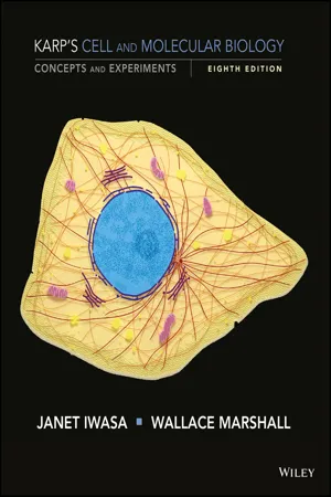
Karp's Cell and Molecular Biology
Concepts and Experiments
- Gerald Karp, Janet Iwasa, Wallace Marshall(Authors)
- 2016(Publication Date)
- Wiley(Publisher)
Mitosis is a process of nuclear division in which the replicated DNA molecules of each chromosome are faithfully segregated into two nuclei. Mitosis is usually accompanied by cytokinesis, a process by which a dividing cell splits in two, partitioning the cytoplasm into two cellular packages. The two daughter cells resulting from Mitosis and cytokinesis possess a genetic content identical to each other and to the mother cell from which they arose. Mitosis, therefore, main- tains the chromosome number and generates new cells for the growth and maintenance of an organism. Mitosis can take place in either haploid or diploid cells. Haploid mitotic cells are found in fungi, plant gametophytes, and a few animals (including male bees known as drones). Mitosis is a stage of the cell cycle when the cell devotes virtually all of its energy to a single activity—chromosome segregation. As a result, most metabolic activities of the cell, includ- ing transcription and translation, are curtailed during Mitosis, and the cell becomes relatively unresponsive to external stimuli. We have seen in previous chapters how much can be learned about the factors responsible for a particular process by studying that process outside of a living cell (page 263). Our understanding of the biochemistry of Mitosis has been greatly aided by the use of extracts prepared from frog eggs. These extracts contain stockpiles of all the materials (histones, tubulin, etc.) necessary to support Mitosis. When chromatin or whole nuclei are added to the egg extract, the chromatin is compacted into mitotic chromosomes, which are segregated by a mitotic spindle that assembles spontane- ously within the cell‐free mixture. In many experiments, the role of a particular protein in Mitosis can be studied by removing that pro- tein from the egg extract by addition of an antibody (immunodeple- tion) and determining whether the process can continue in the absence of that substance (see Figure 14.21 for an example). eBook - PDF
- Ram J. Singh(Author)
- 2016(Publication Date)
- CRC Press(Publisher)
85 4 Cell Division 4.1 INTRODUCTION Cell division is a continuous process that occurs in all living organisms. It has been divided into two categories: Mitosis and meiosis. Both forms of nuclear division occur in eukaryotes and these pro-cesses comprise the cell cycle: G 1 (growth) → S (synthesis of DNA) → G 2 (growth) → M (Mitosis or meiosis) → C (cytokinesis) (Smith and Kindfield, 1999). Mitosis occurs in somatic tissues where each chromosome is divided identically into halves, both qualitatively and quantitatively, producing genetically identical to the parent nucleus. In contrast, meiosis takes place in germ cells with the consequence that nuclei with haploid chromosome numbers are produced. Both types of cell divi-sion play an important role in the development and hereditary continuity of a eukaryotic organism. 4.2 Mitosis 4.2.1 P ROCESS OF M ITOSIS The term Mitosis is derived from the Greek word mitos for thread; coined by Flemming in 1879 (see Chapter 1 ). The synonym of Mitosis is karyokinesis, that is, the actual division of a nucleus into two identical parental daughter nuclei. It is also known as equational division because the exact longitudinal division of each chromosome into identical chromatids and their precise distribution into daughter nuclei leads to the formation of two cells; identical to the original cell from which they were derived. The process of mitotic cell division has been divided into six stages: (1) interphase, (2) prophase, (3) metaphase, (4) anaphase, (5) telophase, and (6) cytokinesis. 4.2.1.1 Interphase Two more terms, resting stage and metabolic stage, have been used to identify interphase cells. However, interphase cells should not be described as being in a “resting stage” because their nuclei are very active as they prepare for cell division. The DNA replication and transcription occur during interphase (Manuelidis, 1990). eBook - ePub
- Ram J. Singh(Author)
- 2016(Publication Date)
- CRC Press(Publisher)
4 Cell Division 4.1 INTRODUCTIONCell division is a continuous process that occurs in all living organisms. It has been divided into two categories: Mitosis and meiosis. Both forms of nuclear division occur in eukaryotes and these processes comprise the cell cycle: G1 (growth) → S (synthesis of DNA) → G2 (growth) → M (Mitosis or meiosis) → C (cytokinesis) (Smith and Kindfield, 1999). Mitosis occurs in somatic tissues where each chromosome is divided identically into halves, both qualitatively and quantitatively, producing genetically identical to the parent nucleus. In contrast, meiosis takes place in germ cells with the consequence that nuclei with haploid chromosome numbers are produced. Both types of cell division play an important role in the development and hereditary continuity of a eukaryotic organism.4.2 Mitosis4.2.1 PROCESS OF MITOSISThe term Mitosis is derived from the Greek word mitos for thread; coined by Flemming in 1879 (see Chapter 1 ). The synonym of Mitosis is karyokinesis, that is, the actual division of a nucleus into two identical parental daughter nuclei. It is also known as equational division because the exact longitudinal division of each chromosome into identical chromatids and their precise distribution into daughter nuclei leads to the formation of two cells; identical to the original cell from which they were derived.The process of mitotic cell division has been divided into six stages: (1) interphase, (2) prophase, (3) metaphase, (4) anaphase, (5) telophase, and (6) cytokinesis. 4.2.1.1 InterphaseTwo more terms, resting stage and metabolic stage, have been used to identify interphase cells. However, interphase cells should not be described as being in a “resting stage” because their nuclei are very active as they prepare for cell division. The DNA replication and transcription occur during interphase (Manuelidis, 1990). Interphase consists of three phases: G1 (gap 1; pre-DNA synthesis) phase, S phase (DNA synthesis), and G2 (gap 2; post-DNA synthesis). The duration of mitotic division is short compared to time required for the cells going through interphase (Figure 4.1 ). Thus, “metabolic stage” is a more appropriate term for the interphase cells. The interphase nucleus contains one or more prominent nucleoli and numerous chromocenters depending on the heterochromatic nature of the chromosomes. Chromosomes cannot be traced individually and they are very lightly stained (Figure 4.2a eBook - PDF
- Emea, A(Authors)
- 2018(Publication Date)
- Agri Horti Press(Publisher)
Cell Divisions 1 C HAPTER 1 Cell Divisions Cell division is the process that cells go through in order to divide. Cells may divide for several reasons, and there are two types of cell division depending on the purpose. The cell division associated with sexual reproduction is one type, called meiosis. The other type, the cell division associated with growth and cell replacement or repair, is called Mitosis. In both types of cell division, the nucleus splits and DNA is replicated. The cell division called Mitosis produces daughter cells that have all the genetic material of the parent cell — a complete set of chromosomes. However, chromosomes are not the only material that needs to be divided and transferred to the daughter cells: there are cytoplasm and the cell membrane to divide as well. Cytokinesis is the process of dividing the cytoplasm and the cell membrane, and this process may follow immediately after Mitosis or occur separately, depending on the organism involved. Together, these two processes make up the mitotic phases of the cell cycle. The phases of cell division are prophase, metaphase, anaphase, and telophase, and these occur in both Mitosis and meiosis. A fifth phases called prometaphase occurring between prophase and metaphase is designated by some, but not all sources. Interphase, which is not part of Mitosis, is a preparatory stage during which the parent cell makes a copy of its genetic material so that each daughter cell can have a complete set. Therefore, Mitosis is an ongoing and repetitive process, alternating with interphase. Cell division is the complex phenomenon by which cellular material is divided equally between daughter cells. This process is the final, and microscopically visible, phase of an underlying change that has occurred at molecular and biochemical levels. Before the cell divides by Mitosis, its fundamental components have duplicated-particularly those involved in hereditary transmission. eBook - PDF
Molecular Cytology V1
The Cell Cycle
- Jean Brachet(Author)
- 2012(Publication Date)
- Academic Press(Publisher)
CHAPTER 5 CELL DIVISION I. GENERAL BACKGROUND There are two characteristics of cell division that sharply distinguish living organisms from nonliving machines: reproduction and heredity. When cells di-vide, the two daughter cells have the same heredity because they have received identical DNA molecules (both in amount and in sequence organization). In culture, they will give rise to identical strains until mutations and DNA rear-rangements induce the inevitable diversification that characterizes all living organisms. Finally, cell division remains the only method of reproduction for all asexual organisms. Sexual reproduction will be touched upon when we discuss oocytes and eggs in Volume 2, Chapter 2. In Biochemical Cytology, Chapter 5 was entitled Mitosis, not Cell Division. This change is due to the fact that, in 1957, little was known of the existence of a cell cycle. Although this idea had been proposed as early as 1953 by Howard and Pelc, it had little immediate impact on cytological research and thinking. The progress in this field in the years that followed was the subject of a book by Prescott (1976); a review by Hochhauser et al. (1981) includes about 1200 references on the subject. Today we know that the cell prepares for mitotic division by a series of complex events that ultimately lead to a revolution: Mitosis, which produces two (in theory, at least) genetically identical daughter cells. We may call this a revolution because it is accompanied by a complete rearrangement of almost all of the cell constituents that have been considered so far. The preparatory events that culminate in Mitosis are dominated by DNA replication. This phase of DNA synthesis (Fig. 1) is called the S phase of the cell cycle. Prior to DNA synthesis is the G! phase, which lasts for a variable length of time (it is even absent during the cleavage of fertilized eggs in most animal species). eBook - PDF
Human Heredity
Principles and Issues
- Michael Cummings(Author)
- 2015(Publication Date)
- Cengage Learning EMEA(Publisher)
In Mitosis, one diploid cell divides to form two diploid cells. Each cell has an exact copy of the genetic information con-tained in the parental cell. 2-5 Mitosis Is Essential for Growth and Cell Replacement ■ Human cells are genetically programmed to divide about 50 times. This limit allows growth to adulthood and re-pairs such as wound healing. Alterations in this program can lead to genetic disorders of pre-mature aging or to cancer. 2-6 Cell Division by Meiosis: The Basis of Sex ■ Meiosis is a form of cell division that produces haploid cells containing only one copy of each chromosome. In an early stage of meiosis, members of a chromosome pair physi-cally associate. At this time, each chromosome consists of two sister chromatids joined by a common centromere. In metaphase I, pairs of homologous chromosomes line up at the equator of the cell. In anaphase I, members of a chro-mosome pair separate from each other. Meiosis I produces cells that contain one member of each chromosome pair. In meiosis II, the unpaired chromosomes line up at the middle of the cell. In anaphase II, the centromeres divide, and the daughter chromosomes move to opposite poles. The four cells produced in meiosis contain the haploid number (23 in humans) of chromosomes. 2-7 Formation of Gametes ■ In males, cells in the testes (spermatagonia) divide by mi-tosis to produce spermatocytes, which undergo meiosis to form spermatids. Spermatids undergo structural changes to convert them into functional sperm. In females, ovarian cells (oogonia) divide by Mitosis to form primary oocytes. The primary oocytes undergo meio-sis. In female meiosis, division of the cytoplasm is unequal, leading to the formation of one functional gamete and three smaller cells known as polar bodies. AP Photo/Gerald Herbert Cell Structure Reflects Function 1. What advantages are there in having the interior of the cell divided into a number of compartments such as the nucleus, the ER, lysosomes, and so forth? 2. eBook - PDF
- Cecie Starr, Christine Evers, Lisa Starr, , Cecie Starr, Cecie Starr, Christine Evers, Lisa Starr(Authors)
- 2020(Publication Date)
- Cengage Learning EMEA(Publisher)
● ● Division of a eukaryotic cell occurs in two steps: nuclear division followed by cytoplasmic division. ● ● Mitosis, the nuclear division mechanism that maintains the chromosome number, is the basis of growth, tissue repair, and (in many species) asexual reproduction. 9.3 Mitosis and Cytoplasmic Division LEARNING OBJECTIVES ●●● ● Describe the role of microtubules in nuclear division. ●●● ● Explain the difference between cytoplasmic division in plants and animals. Stages of Mitosis When a cell is in interphase, its chromosomes are loosely packed to allow transcrip- tion and DNA replication (Figure 9.5). Loose packing makes chromosomes difficult to see under a light microscope 1 . The events of Mitosis proceed in four main stages: prophase, metaphase, ana- phase, and telophase. Prophase Mitosis begins with prophase. During prophase, the chromosomes pack into their most compact forms, which are visible under a light microscope 2 . Tight packing keeps the chromosomes from getting tangled and breaking as the nucleus divides. “Mitosis” is from mitos, the Greek word for “thread,” after the threadlike appearance of chromosomes during the process. The nuclear envelope breaks up as microtubules lengthen from two regions on opposite sides of the cell and attach to the chromosomes at their centromeres. The microtubules form a spindle, a temporary structure that moves chromosomes during nuclear division (Figure 9.6). The areas where the spindle originates on both sides of the cell are called spindle poles. Metaphase By the end of prophase, one sister chromatid of each chromosome has become attached to microtubules extending from one spindle pole, and the other sister chromatid has become attached to microtubules extending from the other spindle pole. The opposing sets of microtubules then begin a tug-of-war. By adding and losing tubu- lin subunits, the microtubules lengthen and shorten, pushing and pulling the chromo- somes as they do. eBook - PDF
- John H. Lawrence, Cornelius A. Tobias, John H. Lawrence, Cornelius A. Tobias(Authors)
- 2013(Publication Date)
- Academic Press(Publisher)
The situation in bacteria remains unresolved. Bodies staining like chromosomes and aligning themselves in the dividing cell in arrangements similar to mitotic figures have been described by DeLamater and others (25, 26), working with painstaking techniques at the very limits of resolution of the light microscope. These observations have not gone unchallenged (6). It is now established in a considerable number of cases that the actual duplication of the chromosomes takes place between divisions or, at the latest, early in the prophase (40, 109, 113). Therefore, the essential fea-ture of Mitosis is the separation of the daughters produced by the duplica-tion of a set of chromosomes in such a way that one of each pair of daughters goes to each daughter nucleus. In the typical plant and animal case, the nuclear membrane disappears, and the process of separating 72 DANIEL MAZIA TABLE I Major Variations of the Scheme of Cell Division I. Mitosis leading to the formation of separate daughter nuclei. A. Nuclear behavior during prophase. 1. Nuclear membrane disappears (most plants and animals). 2. Nuclear membrane persists through division (many protozoa, some animals). B. Chromosome alignment at metaphase. 1. All of chromosomes contained in metaphase plate (many large cells with small chromosomes). 2. Kinetochores (centromeres) only aligned on equatorial plane. Arms of chromosomes dangle from equatorial plane (many small cells with large chromosomes). 3. Equatorial alignment not evident. Chromosomes may lie entirely outside continuous spindle, connected to poles by chromosomal fibers. Metaphase plate may lie obliquely to axis of spindle (pollen tubes). C. Spindle form. 1. Continuous spindle fibers from pole to pole intermingled with chromosomal fibers connecting chromosomes to poles (many plant and animal cells). 2. Continuous central spindle may be distinct from half spindles con-necting chromosomes to poles. eBook - PDF
Cells and Tissues in Culture
Methods, Biology and Physiology
- E. N. Willmer(Author)
- 2015(Publication Date)
- Academic Press(Publisher)
These 204 H. FIRKET reviews are often longer than the present chapter could reasonably be, so the latter is bound to be very incomplete. Most of the older literature was reviewed in Hughes' book The Mitotic Cycle (1952) and the very large and recent treatise by Mazia (1961) covers an immense and varied ground. In this chapter we cannot completely exclude discussion of various cells whose behaviour may be rather different from that of animal tissue cells; it would be rather arbitrary to do so. But, in centering on cultured somatic vertebrate cells, the homogeneity of the material is increased and the problems may perhaps be better cir-cumscribed. As in all other fields of Cell Biology, the trends have been towards a more physiological approach and an evaluation of chemical events. The electron-microscope has made less impact on this than it has in other chapters of Cytology. For the first time, the question of the mechanism of Mitosis has begun to emerge from the domain of speculation. The most important point acquired about cell division in the last decade is that it does not consist of a rapid duplication of cell elements, but essentially of an equal separation of parts previously synthesized. Indeed, Mitosis could be said to be mainly movement and catabolism, the necessary anabolic part of cell multiplication having occurred earlier. This is found to be true of all parts and constituents of the cell, as technical advances pro-gressively permit their separate study. This justifies the amount of space devoted in this chapter to events occurring before Mitosis. II. Mitosis A. GENERAL SURVEY 1. Definition of Phases It is not necessary here to give again at length the classical description of Mitosis, familiar to every reader. Observation of living cells by phase-contrast has brought about a habit of subdividing Mitosis into 5 phases, separated by sudden morphological changes—not always so easy to identify on fixed cells—rather than the old-time 4 stages. eBook - PDF
- Gerald Karp, Janet Iwasa, Wallace Marshall(Authors)
- 2018(Publication Date)
- Wiley(Publisher)
L. Samuels, T. H. Giddings, Jr., & L. A. Staehelin, Journal Cell Biology 130:1354, 1995. The Journal of Cell Biology by Rockefeller Institute; American Society for Cell Biology. Copyright 1995 reproduced with permission of Rockefeller University Press in the format republish in a textbook via copyright clearance center. 3 2 1 Parent cell wall Plasma membrane Microtubule _ + Vesicle (b) (a) 608 CHAPTER 14 • Cell Division 14.11 Meiosis The production of offspring by sexual reproduction includes the union of two cells, each with a haploid set of chromosomes. As discussed in Chapter 4, the doubling of the chromosome number at fertilization is compensated by an equivalent reduction in chromosome number at a stage prior to formation of the gam- etes. This is accomplished by meiosis, a term coined in 1905 from the Greek word meaning “reduction.” Meiosis ensures pro- duction of a haploid phase in the life cycle, and fertilization ensures a diploid phase. Without meiosis, the chromosome number would double with each generation, and sexual repro- duction would not be possible. To compare the events of Mitosis and meiosis, we need to examine the fate of the chromatids. Prior to both Mitosis and meiosis, diploid G 2 cells contain pairs of homologous chromo- somes, with each chromosome consisting of two chromatids. During Mitosis, the chromatids of each chromosome are split apart and separate into two daughter nuclei in a single division. As a result, cells produced by Mitosis contain pairs of homolo- gous chromosomes and are genetically identical to their par- ents. During meiosis, in contrast, the four chromatids of a pair of replicated homologous chromosomes are distributed among four daughter nuclei. Meiosis accomplishes this feat by incor- porating two sequential divisions without an intervening round of DNA replication (FIGURE 14.39). In the first meiotic divi- sion, each chromosome (consisting of two chromatids) is sepa- rated from its homologue. eBook - ePub
- Julia E. Richards, R. Scott Hawley(Authors)
- 2010(Publication Date)
- Academic Press(Publisher)
During the next step, S, DNA synthesis copies the chromosomes. During G2, the cell finishes off any remaining metabolic processes needed for cell division. Interphase consists of G1 plus S plus G2, a period during which the cell looks pretty much the same under the microscope. The microscopic view starts to change during M phase, or Mitosis, when the chromosomes condense (prophase), line up (metaphase), move to the separate ends of the dividing cell (anaphase), and then are packaged back into a nucleus as the cell prepares to divide (telophase) in an orderly manner. Cytokinesis divides the cell into the two new daughter cells. Cells that are growing slowly spend a lot of time in G1. This pie chart shows an average representation of amount of time in the cell cycle spent in each of these stages. It also shows that Mitosis is a very brief part of the cell cycle. If cells are truly inactive and not dividing, they go into a metabolic resting state called G0 instead of going into G1. Take-home message: During interphase, when the nucleus looks like a Brillo pad, the cell makes copies of everything and gets ready for cell division. During the visibly distinct stages of Mitosis, the cell carts the chromosomes around to where they should be (a process we can see under the microscope), and cytokinesis completes the separation into two cells. Through most of the cell’s life the DNA molecules are loosely entwined with each other in the cell nucleus, going about the gentle business of running various aspects of metabolism and growth through transcription of genes to produce functional gene products. During this time, the chromosomes are not visible as separate entities; rather, the nucleus looks like an old Brillo pad. Only once the cell starts the process of Mitosis do we begin to see distinct structures within the nucleus. So let’s take a look at Mitosis and see how it works. 6.2 eBook - PDF
- Donald Voet, Judith G. Voet(Authors)
- 2023(Publication Date)
- Wiley(Publisher)
b. Germ Cells Are Formed by Meiosis The formation of germ cells, a process known as meiosis (Fig. 1-22), requires two consecutive cell divisions. Before the first meiotic division each chromosome replicates, but the resulting sister chromatids remain attached at their cen- tromere. The homologous pairs of the doubled chromosomes then line up across the equatorial plane of the cell in zipper- like fashion, which permits an exchange of the corresponding sections of homologous chromosomes in a process known as crossing-over. The spindle then moves the members of Figure 1-20 Chromosomes. A photomicrograph of a plant cell (Scadoxus katherinae Bak.) during anaphase of Mitosis showing its chromosomes being pulled to opposite poles of the cell by the mitotic spindle. The microtubules forming the mitotic spindle are stained red and the chromosomes are blue. [Courtesy of Andrew S. Bajer, University of Oregon.] Figure 1-21 Mitosis, the usual form of cell division in eukaryotes. Mitosis yields two daughter cells, each with the same chromosomal complement as the parental cell. Section 1-4 Genetics: An Overview 25 each homologous pair to opposite poles of the cell so that, after the first meiotic division, each daughter cell contains N doubled chromosomes. In the second meiotic division, the sister chromatids separate to form chromosomes and move to opposite poles of the dividing cell to yield a total of four haploid cells that are known as gametes. Fertilization consists of the fusion of a male gamete (sperm) with a female gam- ete (ovum) to yield a diploid cell known as a zygote that has received N chromosomes from each of its parents. B. Mendelian Inheritance The basic laws of inheritance were reported in 1866 by Gregor Mendel.
Index pages curate the most relevant extracts from our library of academic textbooks. They’ve been created using an in-house natural language model (NLM), each adding context and meaning to key research topics.