Biological Sciences
Prokaryotes and Viruses
Prokaryotes are single-celled organisms that lack a nucleus and other membrane-bound organelles. They include bacteria and archaea. Viruses are non-living infectious agents that can only replicate inside the cells of living organisms. They consist of genetic material (DNA or RNA) surrounded by a protein coat. Both prokaryotes and viruses play significant roles in various ecological and biological processes.
Written by Perlego with AI-assistance
Related key terms
1 of 5
10 Key excerpts on "Prokaryotes and Viruses"
 No longer available |Learn more
- Laurie Ann Callihan, David Callihan, David Callihan(Authors)
- 2013(Publication Date)
- Research & Education Association(Publisher)
Many species have only a single cell, others are multicellular. (Although viruses are sometimes considered to be living, they are non-cellular and cannot fulfill the characteristics of life without invading the cell of another organism.) Cell structure varies according to the function of the cell and the type of living thing. There are two main types of cells: prokaryotic and eukaryotic. Prokaryotes have no nucleus or any other membrane-bound organelles (cell components that perform particular functions). The DNA in prokaryotic cells usually forms a single chromosome, which floats within the cytoplasm. Prokaryotic organisms have only one cell and include all bacteria. Plant, fungi, and animal cells, as well as protozoa, are eukaryotic. Eukaryotic cells contain membrane-bound intracellular organelles, including a nucleus. The DNA within eukaryotes is organized into chromosomes. A single organism can be unicellular (consisting of just one cell), or multicellular (consisting of many cells). A multicellular organism may have many different types of cells that differ in structure to serve different functions. Individual cells may contain organelles that assist them with specialized functions. For example, muscle cells tend to contain more mitochondria (organelles that make energy available to the cells) since muscle requires the use of extra energy. Animal cells differ in structure and function from photosynthetic cells, which are found in plants, some bacteria, and some protists. Photosynthetic cells have the added job of producing food so they are equipped with specialized photosynthetic organelles. Plant cells also have a central vacuole and cell walls, structures not found in animal cells. Fig. 3-1 Viruses and Cells. Viruses are much smaller than cells, ranging from approximately 0.05-0.1 micrometers. The prokaryotic cell has no nucleus or other membrane-bound organelles and is approximately 1-10 micrometers in diameter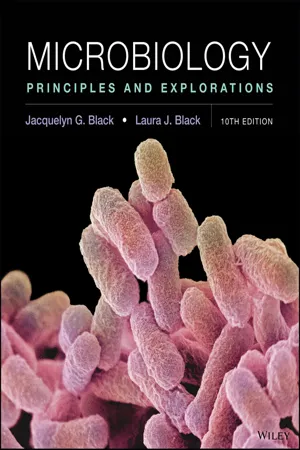 eBook - PDF
Microbiology
Principles and Explorations
- Jacquelyn G. Black, Laura J. Black(Authors)
- 2018(Publication Date)
- Wiley(Publisher)
However, some viruses infect prokaryotic cells, while other viruses infect eukaryotes. ▶ Chapter 11 will examine viruses in detail. PROKARYOTIC CELLS Detailed studies of cells have revealed that prokaryotes differ enough to be split into two large groups called domains. A relatively new concept in biological classifi- cation, domain is the highest category, higher even than kingdom. Three domains exist: two prokaryotic and one eukaryotic: • Archaea (archaeobacteria) (from archae, ancient) • Bacteria (eubacteria) • Eukarya All members of Archaea and Bacteria are prokary- otes and have traditionally been called types of bacteria. A problem of terminology arises over the use of a capi- tal versus a lowercase b in the word bacteria. All bacteria (lowercase b) are prokaryotes, but not all prokaryotes belong to the domain Bacteria (capital B). The differ- ences between Archaea and Bacteria are not so much structural as molecular. Therefore, most of what we have to say about “bacteria” in this chapter applies to both Archaea and Bacteria. (We will discuss Archaea further in ▶ Chapter 12.) Most bacteria on this planet, both in the environment and living in and on humans, are members of the domain Bacteria. As yet, we know of no disease-causing Archaea, BASIC CELL TYPES All living cells can be classified as either prokaryotic, from the Greek words pro (before) and karyon (nucleus), or eukaryotic, from eu (true) and karyon (nucleus). Pro- karyotic (pro-kar″e-ot′ik) cells lack a nucleus and other membrane-enclosed structures, whereas eukaryotic (u-kar″e-ot′ik) cells have such structures. All prokaryotes are single-celled organisms, and all are bacteria. Most of this book will be devoted to the study of prokaryotes. Eukaryotes include all plants, animals, fungi, and protists (organisms such as Amoeba, Paramecium, and the malaria parasite). eBook - PDF
- Rodney P. Anderson, Linda Young, Kim R. Finer(Authors)
- 2020(Publication Date)
- Wiley(Publisher)
It will also summarize how prokaryotes evolved, how they are clas- sified and how they have changed the chemical and physical makeup of the planet. Prokaryotic Organisms The Staphylococcus aureus bacteria growing on the cilia of human nasal epithelial cells make up part of our personal prokaryotic world. J. Berger/Science Source CHAPTER 5 95 96 CHAPTER 5 Prokaryotic Organisms TABLE 5.1 A Comparison of Prokaryotic and Eukaryotic Cells Feature Prokaryotes Eukaryotes Size 0.2–5 μm 10–100+ μm Genetic material Genetic information carried by DNA in one nucleoid or circular chromosome; may contain plasmids carrying accessory genes Genetic information carried by DNA in several linear chromosomes Protein synthesis 70S ribosomes 80S ribosomes Cell wall Structurally complex, composed of peptidoglycan Absent or structurally simple, composed of cellulose, chitin, and silica DNA organization No membrane-bound nucleus True membrane-bound nucleus Organelles No true membrane-bound organelles Many complex membrane-bound organelles Cell division Replicate by binary fission, a simple cell division process that separates copied nucleoids into daughter cells Replicate by mitosis/meiosis, complex processes to precisely separate many copied chromosomes into daughter cells Organism composition Unicellular bacteria and archea Mostly unicellular protists and multicellular plants, fungi, and animals Example of a single-celled organism The bacterium Escherichia coli The alga Euglena gracilus 5.1 The Prokaryote’s Place in the Living World LEARNING OBJECTIVES 1. Describe the roles of prokaryotes in sustaining life. 2. Explain the symbiotic interactions prokaryotes can have with other living organisms. Prokaryotes are small, unicellular organisms that lack a distinct nucleus and complex membrane-bound organelles. Their cellular makeup is distinctly different from the cells of eukaryotes—the microscopic protozoans and the plants, ani- mals, and fungi of the macroscopic world (Table 5.1). eBook - PDF
- Joyce James, Colin Baker, Helen Swain(Authors)
- 2008(Publication Date)
- Wiley-Blackwell(Publisher)
5.1 Eukaryotic and prokaryotic cell structure. them from antibiotics and being attacked by white blood cells. Bacteria working together like this is a bit like an organ system; different types carry out different functions for their mutual benefit so that all of them can survive in hostile environments. Viruses and Prions Viruses and prions are not cells. They can be thought of as being somewhere between the living and the dead! However they depend on cells for their replication. Nearly all of them are smaller than the smallest cell (Table 5.1). Viruses have either ribonucleic acid (RNA) or deoxyribonucleic acid (DNA) as their genetic material; they can survive outside their host cell in an extracellular state. In the extracellular state, a protein coat and sometimes a mem-branous envelope surrounds the genetic mater-ial. In this state they are called virus particles or virions. Prions are made entirely from glycoproteins and have no nucleic acids. Like viruses they have an extracellular state but little is known about prions. When cells are infected with a virus or prion, it is usually damaging to the health of the infected organism, e.g. influenza and mad cow Activity Put some Sellotape on the back of your hand and peel it off. Hold the tape to the light and see the outer skin cells. Activity Look at a bacterial biofilm by scraping some plaque from your teeth. If you can, look at it under a microscope. You can also see the biofilm by using disclosing tablets on your teeth. disease (Creutzfeldt-Jakob syndrome) caused by a virus and a prion respectively. Cell structure The small size of cells means that the detailed structure of cells and viruses can only be seen with an electron microscope which can magnify objects about 100 000 times. An electron microscope can be used diagnostically, e.g. ex-amination of infant faecal samples for human rotavirus. Electron microscopes are very big and expen-sive and not usually found in a basic hospital laboratory. eBook - PDF
- Fitch, J. Patrick(Authors)
- 2002(Publication Date)
At this point it is essential to introduce the vocabulary needed to discuss living organisms. We have focused the discussion on the topics needed for working in the biotechnology field. Since the 1930s the living world has been divided into two different domains of organisms called eukaryotes and prokaryotes. Eukaryote translates into “true kernel” or “with nucleus.” Prokaryote translates into “without kernel or nucleus.” We will interchange common use and misuse of the Latin roots and conjugations—e.g., the popular term prokaryotes and the proper Latin plural prokarya . Traditionally, after the domains Eukarya and Prokarya, the next levels 4 Chapter 1 of separation of living organisms are kingdoms, followed by phyla, classes, orders, families, genera, species, and strains. The Eukarya include four kingdoms: Protista, Plantae, Fungi, and Animalia. The Protista are single-celled organisms, such as the protozoa. The Plantae are multicellular organisms that manufacture their food, such as ferns and trees. The Fungi are single or multicellular organisms that absorb food from the environment, such as yeasts and molds. The Animalia are multicellular organisms that must capture food and digest it internally, such as dogs, birds, and fish. Eukaryotes are the organisms that most people would observe in their daily routine and include people, animals, plants, and fungi. The presence of a nucleus and other cellular organelles is specific to Eukarya and complicates understanding how these cells work. Prokarya were traditionally defined as the Monera kingdom and included both bacteria and archaebacteria. In the past two decades, Carl Woese of the University of Illinois and others have proposed the archaea as a separate domain of life—distinct from bacteria. Archaea are often extremophiles—living, for instance, in high salt (halophilic archaea) or high temperature (thermophilic archaea) environments.
- Cecie Starr, Christine Evers, Lisa Starr, , Cecie Starr, Cecie Starr, Christine Evers, Lisa Starr(Authors)
- 2020(Publication Date)
- Cengage Learning EMEA(Publisher)
Editorial review has deemed that any suppressed content does not materially affect the overall learning experience. Cengage Learning reserves the right to remove additional content at any time if subsequent rights restrictions require it. 286 UNIT 3 EVOLUTION AND DIVERSITY 14.7 Viruses LEARNING OBJECTIVES ●●● ● Explain how viruses replicate. ●●● ● Compare the structure of a bacteriophage and the human immunodeficiency virus (HIV). ●●● ● Explain how new combinations of viral genes can arise. Although viruses are not cells, they are considered part of the human microbiota. A virus is a noncellular infectious particle that replicates only inside a living cell. We do not know how viruses are related to cellular life. The fact that they can only replicate inside cells suggests that they may have evolved from cells. Alternatively, viruses may be remnants of a time before cells. Viruses infect and replicate in all organisms, no matter how simple or complex. A viral infection often decreases a host’s ability to survive and reproduce, so viruses affect ecological interactions among species throughout the biosphere. Viral Structure A free viral particle (one that is not inside a cell) always includes a viral genome enclosed within a protein coat. The viral genome may be RNA or DNA, and it may be single-stranded or double-stranded. The viral coat consists of many pro- tein subunits that self-assemble in a repeating pattern to produce a helical rod (Figure 14.32A) or many-sided (polyhedral) structure (Figure 14.32B). The coat pro- tects the viral genetic material and plays a role in infection. In all viruses, compo- nents of the viral coat bind to proteins at the surface of a host cell. The specificity of this binding ensures that each type of virus can infect on only one species or a group of related species. In addition to the viral genome, the coat may also enclose viral enzymes that will act within the host. eBook - PDF
Infrastructure and Activities of Cells
Biotechnology by Open Learning
- M.C.E. van Dam-Mieras, B C Currell, R C E Dam-Mieras(Authors)
- 2016(Publication Date)
- Butterworth-Heinemann(Publisher)
Nucleus, plastids and cytoplasm became part of the biologists vocabulary. The advent of the electron microscope earlier this century with a much greater magnifying power than that of light microscopes, led to greater resolution of the fine structure of cells. These studies clearly demonstrated that cells can be divided into two quite distinct types described as prokaryotic and eukaryotic. Prokaryotic cells are structurally the simpler of the two. This type of cell morphology is confined to the bacteria. The cells of other groups of micro-organisms (eg algae, fungi and protozoa) and of all plants and animals are of the eukaryotic type. Prokaryotic cells are much smaller than eukaryotic cells. Most of those organisms which exhibit prokaryotic organisation are unicellular (ie each organism consists of a single cell) whilst many eukaryotes exhibit multicellularity (ie each organism consists of many cells which may be of many different types). In this chapter, you will learn about the basic structure of prokaryotic cells. Subsequent chapters will deal with eukaryotic cell structure and function. Despite the fact that prokaryotic organisation is restricted to the prokaryotes, it is still important to have a good understanding of how such cells function since bacteria include some important disease causing types as well as potential mediators of many important biotechnological processes. They also include some types which are responsible for major environmental and geochemical changes. The fundamental differences between prokaryotes and eukaryotes has important consequences in medicine, genetic manipulation and biotechnology. The architecture of prokaryotic cells 3 This first chapter is quite long so do not attempt to do it all in one sitting. eBook - PDF
- Mary Ann Clark, Jung Choi, Matthew Douglas(Authors)
- 2018(Publication Date)
- Openstax(Publisher)
The name "prokaryote" suggests that prokaryotes are defined by exclusion—they are not eukaryotes, or organisms whose cells contain a nucleus and other internal membrane-bound organelles. However, all cells have four common structures: the plasma membrane, which functions as a barrier for the cell and separates the cell from its environment; the cytoplasm, a complex solution of organic molecules and salts inside the cell; a double-stranded DNA genome, the informational archive of the cell; and ribosomes, where protein synthesis takes place. Prokaryotes come in Chapter 22 | Prokaryotes: Bacteria and Archaea 595 various shapes, but many fall into three categories: cocci (spherical), bacilli (rod-shaped), and spirilli (spiral- shaped) (Figure 22.9). Figure 22.9 Common prokaryotic cell types. Prokaryotes fall into three basic categories based on their shape, visualized here using scanning electron microscopy: (a) cocci, or spherical (a pair is shown); (b) bacilli, or rod-shaped; and (c) spirilli, or spiral-shaped. (credit a: modification of work by Janice Haney Carr, Dr. Richard Facklam, CDC; credit c: modification of work by Dr. David Cox; scale-bar data from Matt Russell) The Prokaryotic Cell Recall that prokaryotes are unicellular organisms that lack membrane-bound organelles or other internal membrane-bound structures (Figure 22.10). Their chromosome—usually single—consists of a piece of circular, double-stranded DNA located in an area of the cell called the nucleoid. Most prokaryotes have a cell wall outside the plasma membrane. The cell wall functions as a protective layer, and it is responsible for the organism’s shape. Some bacterial species have a capsule outside the cell wall. The capsule enables the organism to attach to surfaces, protects it from dehydration and attack by phagocytic cells, and makes pathogens more resistant to our immune responses. eBook - PDF
- Marc H.V. van Regenmortel, Brian W.J. Mahy(Authors)
- 2010(Publication Date)
- Academic Press(Publisher)
Another major breakthrough in recent viral research was indeed the realization that viruses are much more abundant than cells and much more diverse than previously suspected. It is thus currently assumed that viral genomes represent the major throve of genetic diversity on Earth. All these trends make it irrelevant to ask whether viruses are alive. As recently pointed out by Jean-Michel Claverie, the question of the nature of viruses has been for a long time obscured by the confusion between virus and virus particle, whereas the major components of the virus life cycle correspond to the intracellular viral factory. All this concurs to put again the question of virus origin on the agenda. Thus a brief summary of the main data that presently point to an ancient origin of viruses and the discussion of how the three major hypotheses explaining the origin of viruses have been rejuvenated in this new framework are presented here. Viruses Are Ancient For a long time, virologists thought that the various virus families were evolutionary unrelated, indicating a polyphyletic origin. In recent years, this view has progressively changed with the identification of more and more relationships (sometimes totally unexpected) between different viral lineages, making it possible to define a limited number of large viral groups whose hosts encompass the three cellular domains. For example, it has been clearly established that some double-stranded RNA viruses infecting bacteria are homologous to those infecting eukarya, suggesting that they predated Origin of Viruses 25 the divergence between bacteria and eukaryotes. Since the RNA replicases/transcriptases of these double-stranded RNA viruses are homologous to those of single-stranded RNA viruses, all RNA viruses presently known seem to be evolutionary related (at least in term of their replication apparatus). eBook - PDF
Viruses
A Natural History
- Marilyn J. Roossinck(Author)
- 2023(Publication Date)
- Princeton University Press(Publisher)
VIRUSES IN ECOSYSTEM BALANCE 196 V I R U S E S I N E C O S Y S T E M B A L A N C E In the late 1980s scientists estimated the number of viruses in the sea by adding a milliliter of filtered seawater to a plate of a lab strain of the bacteria Escherichia coli. Bacterial viruses often kill their hosts, and when a virus infects E. coli on a petri dish it makes a little hole in the bacterial lawn where the cells have died. Each hole represents a single infectious virus. Using this method, the researchers calculated that there are about a million viruses that can infect E. coli in a milliliter of seawater. Viruses in the sea This didn’t include any of the viruses that don’t infect E. coli or those that don’t kill their hosts. Later estimates were made using electron microscopy or fluorescent imaging (see page 36), which revealed all the viruses present in a sample of seawater, regardless of their hosts. These put the figure of marine viruses at 10 million virus particles per milliliter of seawater. What are all these viruses doing? Which organisms are they infecting? These are complex questions and we certainly don’t know all the answers yet, but we do know the genome sequences for many marine viruses and we also know that most of them infect microbes. Understanding the details relies on sophisticated computer analyses and comparisons, part of the important field of bioinformatics. A plaque assay is an analysis for viruses that can infect bacteria. The sample is filtered to remove bacteria and then added to a bacterial lawn. When a virus infects a bacterial cell, it kills the cell and spreads to nearby cells, which are also killed. This leaves a small hole, or plaque, in the bacterial lawn, each of which represents a single virus. 197 V I R U S E S I N T H E S E A Microbes are the main contributors to the biomass of the oceans, and most are bacteria. In terms of sheer numbers, however, viruses exceed other microbes by at least tenfold.
Index pages curate the most relevant extracts from our library of academic textbooks. They’ve been created using an in-house natural language model (NLM), each adding context and meaning to key research topics.