Psychology
REM sleep
REM sleep, or rapid eye movement sleep, is a stage of sleep characterized by rapid eye movements, vivid dreams, and heightened brain activity. It is one of the five stages of sleep and is associated with memory consolidation, learning, and emotional processing. During REM sleep, the body experiences temporary paralysis to prevent acting out dreams.
Written by Perlego with AI-assistance
Related key terms
1 of 5
10 Key excerpts on "REM sleep"
 eBook - PDF
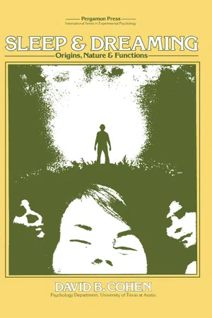
Sleep & Dreaming
Origins, Nature and Functions
- D. Cohen, H. J. Eysenck(Authors)
- 2013(Publication Date)
- Pergamon(Publisher)
And perhaps one psychological function of REM sleep is to provide a biological context within which to process such information. A somewhat different approach to the functional significance of REM sleep focuses on the effect on REM characteristics (e.g., eye movements, REM time) of a novel learning situation. It has been hypothesized that REM sleep promotes the consolidation of new learn-ing (Dewan, 1969; Fishbein and Gutwein, 1977; McGrath and Cohen, 1978; Shapiro, 1967). INFORMATION PROCESSING DURING REM sleep 79 This idea is similar to clinical views on the role of dreaming in the integration of recent and long term memories that are of affective significance (Breger, 1967). The programming hypothesis is readily testable once the assumption is made that certain characteristics of REM (duration, eye movement density, latency) reflect the amount or intensity of, or motivation for, information processing. However, it should be noted that the assumption is still an empirical question. Typically, REM deprivation is used to assess the role of REM sleep in the mediation of learning. A little-used alternate technique is to study the characteristics of REM during uninterrupted sleep following learning. For example, Fishbein and Kastaniotis (1973) reported an increase in REM time over baseline in mice trained for one hour on an active avoidance task compared to yoked controls. Likewise, Lecas (1976) reported that REM but not NREM time increased during sleep subsequent to new learning in cats, an effect that was maximal just prior to reaching criterion. In a preliminary study of six 6-month-old babies it was reported that following successful learning (head turning response reinforced by visual stimulation), REM time but not quiet sleep time increased over control sessions (Paul and Dittrichova, 1975). In addition, there was no increase in REM time during sleep which followed unsuccessful learning (which required a double head turn, too difficult for the babies to learn).
- Kelly Bulkeley Ph.D.(Author)
- 2017(Publication Date)
- Praeger(Publisher)
Every clinician seemed to come up with a different theory about the nature of dreaming. Alternative models, methods, and approaches multiplied rapidly. Rather than moving toward greater clarity and consensus, dream psychologists appeared to be mov- ing further and further apart, with their theories increasingly contradict- ing one another. A dramatic change in the history of 20th-century dream psychology occurred in the 1950s when sleep laboratory researchers discovered the phenomenon of rapid eye movement (REM) sleep and its associations with dreaming. Suddenly the possibility opened up that dreaming could be objectively observed, precisely measured, and experimentally studied in a controlled laboratory situation. At long last, it appeared a truly scientific psychology of dreaming could be developed. This chapter will describe the original research on REM sleep and will discuss how the subsequent explosion of laboratory investigations of REM sleep has transformed the modern psychology of dreaming. Sleep Laboratories, REM sleep, and Dreaming 61 Aserinsky and Kleitman’s Discovery of REM sleep Nathaniel Kleitman was a physiologist at the University of Chicago who had spent many years studying the nature of sleep, particularly the effects of sleep deprivation. One of his students was Eugene Aserinsky, who was doing graduate work investigating aspects of attention in chil- dren. While collecting data for his project, Aserinsky noticed one day that the children’s eyes began shifting around very quickly beneath their lids whenever they lost attention and fell asleep. These darting eye movements were completely different from the slow, rolling eye movements that researchers had long observed occurring at sleep onset. Aserinsky men- tioned his observations to Kleitman, and when they began more careful study of the children, they were surprised to find that these eye move- ments were very much like a person’s eye movements while awake. eBook - PDF
- Oleg V. Khlevniuk, Vadim A. Staklo(Authors)
- 1983(Publication Date)
- Yale University Press(Publisher)
Health reflects and ultimately depends upon satisfactory synchronization of the entire array of the body's functional rhythms. This synchronization is accomplished within the brain, which has its own intrinsically generated rhythms that are likewise subject to influence by internal bodily and externally imposed stim-uli and demands. The sleep-wake cycle reflects one of the major functional rhythms of the brain, and as noted in the first epigraph, the REM dreaming phase of sleep has been established as a third major brain-mind state—a window of opportunity for inquiry into mind/brain questions. With the use of modern electroencephalo-graph and polygraph instrumentation, it is possible to identify two alternating major stages of sleep: rapid eye movement (REM) and non-REM sleep. There are four depth levels of non-dreaming sleep (NREM) that are qualitatively similar. Typical visual dreaming oc-curs during the REM or dream periods. Thus the sleep phase of the circadian (twenty-four-hour) sleep-wake cycle contains a REM-NREM ultradian (less-than-twenty-four-hour) cycle. Kleitman (1963) regarded the latter as representing a basic rest-activity cycle. REM periods occur at approximately ninety-minute intervals. Usu-ally there are four or five REM periods of roughly twenty minutes' duration during each night's sleep, usually shorter earlier and lon-ger later in the sleep period. The ninety-minute ultradian rhythm seen during sleep may continue during waking hours, but with quite different psychologic and behavioral manifestations such as movement, eating, drinking, and smoking (Friedman and Fisher 129 BRAIN, COGNITIVE NEUROSCIENCE: THE SECOND KEY 1967). The period of this ninety-minute ultradian rhythm lies be-tween the much shorter ones represented by cardiac rate (approxi-mately one second) and respiration (approximately four seconds) and the much longer ones (twenty-four hours to thirty or ninety days) to be mentioned below.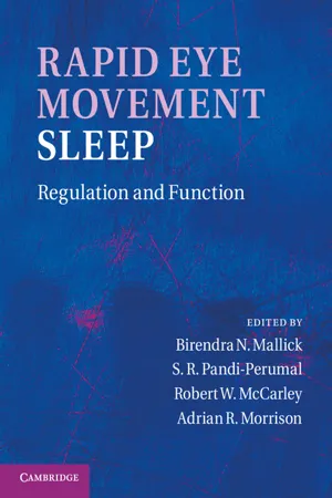 eBook - PDF
Rapid Eye Movement Sleep
Regulation and Function
- Birendra N. Mallick, S. R. Pandi-Perumal, Robert W. McCarley, Adrian R. Morrison, S. R. Pandi-Perumal(Authors)
- 2011(Publication Date)
- Cambridge University Press(Publisher)
Neurosci Biobehav Rev 14(1): 49–63. Wagner, U., Gais, S. & Born, J. (2001) Emotional memory formation is enhanced across sleep intervals with high amounts of rapid eye movement sleep. Learn Mem 8(2): 112–19. Wagner, U., Fischer, S. & Born, J. (2002) Changes in emotional responses to aversive pictures across periods rich in slow-wave sleep versus rapid eye movement sleep. Psychosom Med 64(4): 627–34. Wagner, U., Degirmenci, M., Drosopoulos, S., Perras, B. & Born, J. (2005) Effects of cortisol suppression on known-risk and ambiguous-risk decisions. J Sleep Res 16(3): 245–52. McNamara, P. (1996) REM sleep: a social bonding mechanism. New Ideas Psychol 4(1): 35–46. Mellman, T. A., Bustamante, V., Fins, A. I., Pigeon W. R. & Nolan B. (2002) REM sleep and the early development of posttraumatic stress disorder. Am J Psychiatry 159(10): 1696–701. Mignot, E. (2001) A commentary on the neurobiology of the hypocretin/orexin system. Neuropsychopharmacology 25(5 Suppl): S5–13. Monti, J. M. & Monti, D. (2005) Sleep disturbance in schizophrenia. Int Rev Psychiatry 17(4): 247–53. Nielsen, T. & Levin, R. (2007) Nightmares: a new neurocognitive model. Sleep Med Rev 11(4): 295–310. Nishida, M., Pearsall, J., Buckner, R. L. & Walker, M. P. (2009) REM sleep, prefrontal theta, and the consolidation of human emotional memory. Cereb Cortex 19(5): 1158–66. Nofzinger, E. A., Mintun, M. A., Wiseman, M., Kupfer, D. J. & Moore, R. Y. (1997) Forebrain activation in REM sleep: an FDG PET study. Brain Res 770(1/2): 192–201. Pakyurek, M., Gutkovich, Z. & Weintraub, S. (2002) Reduced aggression in two inpatient children with the treatment of their sleep disorder. J Am Acad Child Adolesc Psychiatry 41(9): 1025. Ponz, A., Khatami, R., Poryazova, R. et al. (2010a) Abnormal activity in reward brain circuits in human narcolepsy with cataplexy. Ann Neurol, 67(2), 190–200. Ponz, A., Khatami, R., Poryazova, R. et al. (2010b) Reduced amygdala activity during aversive conditioning in human narcolepsy. eBook - ePub
- Kelly Bulkeley Ph.D.(Author)
- 2017(Publication Date)
- Praeger(Publisher)
But soon a serious problem arose with this exclusively clinical approach to dream psychology. Every clinician seemed to come up with a different theory about the nature of dreaming. Alternative models, methods, and approaches multiplied rapidly. Rather than moving toward greater clarity and consensus, dream psychologists appeared to be moving further and further apart, with their theories increasingly contradicting one another.A dramatic change in the history of 20th-century dream psychology occurred in the 1950s when sleep laboratory researchers discovered the phenomenon of rapid eye movement (REM) sleep and its associations with dreaming. Suddenly the possibility opened up that dreaming could be objectively observed, precisely measured, and experimentally studied in a controlled laboratory situation. At long last, it appeared a truly scientific psychology of dreaming could be developed.This chapter will describe the original research on REM sleep and will discuss how the subsequent explosion of laboratory investigations of REM sleep has transformed the modern psychology of dreaming.Aserinsky and Kleitman’s Discovery of REM sleep
Nathaniel Kleitman was a physiologist at the University of Chicago who had spent many years studying the nature of sleep, particularly the effects of sleep deprivation. One of his students was Eugene Aserinsky, who was doing graduate work investigating aspects of attention in children. While collecting data for his project, Aserinsky noticed one day that the children’s eyes began shifting around very quickly beneath their lids whenever they lost attention and fell asleep. These darting eye movements were completely different from the slow, rolling eye movements that researchers had long observed occurring at sleep onset. Aserinsky mentioned his observations to Kleitman, and when they began more careful study of the children, they were surprised to find that these eye movements were very much like a person’s eye movements while awake. Aserinsky and Kleitman conducted further experiments, and they soon learned that these periods of highly intensified eye movements, which occurred in both children and adults, were accompanied by distinctive brain wave patterns, irregular breathing, and increased heart rates. eBook - ePub
- (Author)
- 2019(Publication Date)
- Academic Press(Publisher)
LaBerge, Greenleaf, & Kedzierski, 1983 ). Despite the strengths of this paradigm, the infrequency with which most people spontaneously experience lucid dreams remains a challenge to observing them under laboratory conditions and collecting datasets with large sample sizes. Accordingly, a main target of research is to develop methods for the reliable induction of lucid dreams.III Neurobiology of Lucid and Non-Lucid Dreaming
As noted above, while dreamlike mental activity can be observed during all sleep stages, REM sleep dreams are particularly vivid and intense. The specific phenomenal characteristics of REM sleep dreaming have been associated with neural activation patterns observed during this state. For example, higher visual areas show strong metabolic activity during REM sleep (Braun et al., 1998 ), which is in line with the visuospatial hallucinations that are the hallmark of REM sleep dreaming (Windt, 2010 ). The amygdala, parahippocampal cortex, medial prefrontal cortex, and anterior cingulate cortex also show increased activity during REM sleep (Braun et al., 1997 ; Maquet et al., 1996 ). These brain areas have all been implicated in emotional processing, mirroring the intense emotions often experienced in dreams. In contrast, areas of the prefrontal cortex, including the dorsolateral prefrontal cortex and frontal pole, and parietal areas, including the inferior parietal lobule and precuneus, show low metabolic rates during REM sleep (Braun et al., 1997 ; Maquet et al., 1996 ). Hypoactivity of these regions, coupled with preserved or increased activity in limbic/paralimbic structures and extrastriate cortices, has been postulated to facilitate a mode of brain function conducive to hallucinatory dream mentation but diminished higher-order consciousness/self-awareness (Hobson, 1999 ; Maquet, 2000 ). In particular, prefrontal deactivations have been postulated to underlie the cognitive deficiencies typical of ordinary dreaming, such as impaired critical thinking, diminished metacognitive ability, and restricted volitional control (Hobson & Pace-Schott, 2002 eBook - ePub
- Robert Stickgold, Matthew P. Walker(Authors)
- 2010(Publication Date)
- Academic Press(Publisher)
The fact that neuronal activity can be traced to molecular markers of plasticity establishes a strong parallel between the cellular and systems levels, offering support for the Hebbian notion that memory requires two consecutive and distinct steps, namely network reactivation for short-term recall and synaptic remodeling for long-term storage. The current results suggest that SWS and REM sleep play distinct and complementary roles in memory consolidation, with memory recall (network reactivation) occurring mainly during SWS and memory storage (plasticity-related gene) sparked off during REM sleep. Such a mechanism fulfills early conceptual notions of a two-step process for memory consolidation during sleep and is in line with evidence that SWS and REM sleep have synergistic effects on human procedural learning and developmental plasticity.Dreams, Waking, and Sleep Mentation
In adult humans, REM sleep is almost always accompanied by dreaming, as can be easily demonstrated by waking experimental subjects during REM sleep and requesting dream reports. During human REM sleep, a selected set of forebrain areas are reactivated, including portions of the hypothalamus, amygdala, septum, and ventral striatum, as well as the anterior cingulate, orbitofrontal, entorhinal, and insular cortices. The dorsolateral prefrontal cortex, however, shows reduced activity. Given the role of prefrontal cortex activity in the executive control of behavior, its reduction during REM sleep has been proposed to underlie the poor logical concatenation and reduced self-awareness of dreaming in comparison with waking.Dreams may have first evolved as a collateral effect of extended REM sleep, a characteristic mammalian trait. Pet owners know that cats and dogs seem to act out dreams during sleep. More controlled evidence of dreaming in nonhuman mammals was obtained by lesions of the brain stem nuclei that promote muscle atonia during REM sleep. Cats with such lesions sleep quietly through SWS, but upon entering REM sleep they become suddenly agitated by vigorous species-specific behaviors, such as meowing and pouncing. How are dreams generated, and what is their biological function?As noted by Sigmund Freud, dreams often involve elements of the experience of the preceding day(s), the day residue. Although high-fidelity memory reactivation must be at the roots of the cognitive functions of sleep and dreams, it does not account entirely for the symbolic complexity that characterizes the oneiric narrative. After all, it is not common to dream about the exact repetition of waking scenes. On the contrary, most human dreams are characterized by the intrusion of illogical elements, leading to unforeseen associations. Human dreams are subjective narratives composed of familiar and unfamiliar beings, things, and places interacting around a self-representation of the dreamer that mostly observes an unfolding plot. Dreams vary in intensity, ranging from confused and faint impressions to complex time-evolving narratives with vivid imagery and surprising turns. Although dreams tend to be dominated by visual images, they can also involve combinations of auditory, olfactory, tactile, gustatory, motor, vestibular, and linguistic modalities. Dreams can sometimes be extremely pleasant or just the opposite, but they are usually characterized by a mix of emotions. Dreams are also hyperassociative, linking characters, places, and actions in bizarre ways. Dreams can also anticipate events of the coming day(s), particularly when subjects undergo extreme anxiety and expectation. A good example is provided by the dreams of students during the night before taking difficult exams; these dreams often contain detailed anticipatory simulations of the expected challenges, in content and/or context. eBook - PDF
- Rene Drucker-Colin(Author)
- 2012(Publication Date)
- Academic Press(Publisher)
Abstract of the Association for the Psychophysiological Study of Sleep, Denver, 1968. A Motivational Function of REM sleep 249 Pearlman, C. Latent learning impaired by REM sleep deprivation. Psychonomic Science, 2S _i 135-136, 1971. Phillips, C. Actions of antidromic pyramidal volleys on single Betz calls in the cat. Quarterly Journal of Experimental Physiology, 44 : 1-25 , 1959. Roffwarg, H. P., Muzio, J. Ν., and Dement, W. C. Ontogenetic development of the human sleep-dream cycle. Science, 152 : 604-619 , 1966. Rossi, G. F., Favale, E., Hara, T., Guisanni, Α., and Sacco, G. Researches of the nervous mechanisms underlying deep sleep in the cat. Archives of Italian Biology, 99 : 280-292 , 1961. Sachar, E., Hellman, L., Roffwarg, Η., Halpern, F., Fukushima, D. and Gallagher, T. Disrupted 24-hour patterns of Cortisol secretion in psychotic depression. Archive s of General Psychiatry, 28: 19-24, 1973. Satinoff, Ε·, Drucker-Colin, R. R. and Hernandez-Peon, R. Paleocortical excitability and sen-sory filtering during REM sleep deprivation. Physiology and Behavior, 7_: 103-106, 1971. Snyder, F. Progress in the new biology of dreaming. American Journal of Psychiatry, 12 2 : 377-390, 1965 . Steiner, S. S. and Ellman, S. J. Relation between REM sleep and intracranial self stimulation. Science, 177 : 1122-1124 , 1972. Stern, W. C. Effects of REM sleep deprivation upon the acquisition and maintainence of learned behavior in the rat. Abstract of the Associa-tion for the Psychophysiological Study of Sleep, Boston, 1969. Vogel, G. W. A review of REM sleep deprivation. Archive s of General Psychiatry , 3 2 : 749-761, 1975 . 250 Gerald W. Vogel Vogel, G. W., Augustine, F., McAbee, R., and Thurmond, A. New findings about how REM sleep depriva-tion improves depression. Unpublished manu-script and read before the Association for the Psychophysiological Study of Sleep, Houston, 1977. eBook - PDF
- Alexander Golbin, Howard Kravitz, Louis G. Keith, Alexander Golbin, Howard Kravitz, Louis G. Keith(Authors)
- 2004(Publication Date)
- Taylor & Francis(Publisher)
(7) The same psychological variables that predispose the subject to renunciation of search—trait anxiety, fixation on obstacles according to Rosenzweig 34 , motivation of avoidance of failures—have a tendency to correlate with REM percentage 8 . (8) REM sleep is increased in students who display a stable emotional tension both before and after an examination, even with its positive outcome (when the stress situation no longer exists) as compared with students who display emotional tension only before an examination 2 . It is possible to suggest that this stable emotional tension, which is not compatible with the actual situation, is similar to neurotic anxiety and represents renunciation of search. Sleep Psychiatry 46 (9) Healthy subjects with normal search activity during wakefulness are characterized by active participation in their own dreams. This active participation correlates with heart rate acceleration and eye movement density in REM sleep 35 . (10) The more characters and descriptive elements that appear in the dream, and the more active the characters and the dreamer himself, the larger the decrease in the scale of unhappiness during the night 36 . It can be suggested that the characters’ activity represents search activity in dreams. One must stress that, in the case of healthy subjects, the decrease of unhappiness after sleep correlates only with the above- mentioned dream variables, not with the sleep physiology. As I see it, this means that, as long as the dream is functionally effective and is able to restore search activity, REM sleep requirement does not increase. It is increased only when dreams are going to lose their restorative capacity, for instance in clinical or pre-clinical disorders (depression and subsyndromal depression, neurotic anxiety, narcolepsy). eBook - PDF
Restless Legs Syndrome
Diagnosis and Treatment
- William G. Ondo(Author)
- 2016(Publication Date)
- CRC Press(Publisher)
Slow wave sleep (SWS), which will be discussed in more detail later, tends to dominate the first third of sleep, whereas REM sleep is most predominant in the last third of sleep. The first REM cycle is typically short; the final cycle of REM tends to be the longest. In terms of arousa-bility, the deepest sleep occurs in the first third of the night, corresponding to SWS (NREM stages III and IV); an individual awakened during these stages of sleep is typically groggy and confused. If awakened during lighter stages of NREM sleep (stages I and II), there is the possibility that the individual may not be aware that he or she had fallen asleep. However, if one were awakened during REM sleep, a person may experience residual sleep paralysis or persistence of dreams intruding into the waking state because paralysis of voluntary muscles and dreams are nor-mal components of REM sleep. Typical behavioral events that are observed following sleep onset, irrespective of type of sleep, include transient amnesia for the occurrence of sleep, fragmentary images, hypnic jerks, and automatic behavior. An individual’s sleep can be recorded via an overnight polysomnogram (PSG). Some of the most commonly measured variables include electro-encephalography (EEG), electrooculography (EOG), electromyography (EMG), electrocardiography, respiration, oxygen saturation, snoring, and body position (Fig. 2). To score the various stages of sleep, an epoch of sleep, or a 30-second 1 polygraphic tracing at a speed of 10 mm/second, is evaluated according to defined criteria set forth in the international classification of sleep disorders (ICSD) (1). In human adults, NREM sleep occupies 75% to 80% of the total sleep time. It is comprised of stages I through IV. Stage I sleep accounts for about 3% to 8% of the total sleep time. It is scored when an alpha rhythm (8–12 Hz) is present in less than half of the epoch.
Index pages curate the most relevant extracts from our library of academic textbooks. They’ve been created using an in-house natural language model (NLM), each adding context and meaning to key research topics.