
eBook - ePub
Prostate Cancer Imaging
An Engineering and Clinical Perspective
Ayman El-Baz, Gyan Pareek, Jasjit S. Suri, Ayman El-Baz, Gyan Pareek, Jasjit S. Suri
This is a test
Condividi libro
- 376 pagine
- English
- ePUB (disponibile sull'app)
- Disponibile su iOS e Android
eBook - ePub
Prostate Cancer Imaging
An Engineering and Clinical Perspective
Ayman El-Baz, Gyan Pareek, Jasjit S. Suri, Ayman El-Baz, Gyan Pareek, Jasjit S. Suri
Dettagli del libro
Anteprima del libro
Indice dei contenuti
Citazioni
Informazioni sul libro
This book covers novel strategies and state of the art approaches for automated non-invasive systems for early prostate cancer diagnosis. Prostate cancer is the most frequently diagnosed malignancy after skin cancer and the second leading cause of cancer related male deaths in the USA after lung cancer. However, early detection of prostate cancer increases chances of patients' survival. Generally, The CAD systems analyze the prostate images in three steps: (i) prostate segmentation; (ii) Prostate description or feature extraction; and (iii) classification of the prostate status.
- Explores all of the latest research and developments in state-of-the art imaging of the prostate from world class experts.
- Contains a comprehensive overview of 2D/3D Shape Modeling for MRI data.
- Presents a detailed examination of automated segmentation of the prostate in 3D imaging.
- Examines Computer-Aided-Diagnosis through automated techniques.
- There will be extensive references at the end of each chapter to enhance further study.
Domande frequenti
Come faccio ad annullare l'abbonamento?
È semplicissimo: basta accedere alla sezione Account nelle Impostazioni e cliccare su "Annulla abbonamento". Dopo la cancellazione, l'abbonamento rimarrà attivo per il periodo rimanente già pagato. Per maggiori informazioni, clicca qui
È possibile scaricare libri? Se sì, come?
Al momento è possibile scaricare tramite l'app tutti i nostri libri ePub mobile-friendly. Anche la maggior parte dei nostri PDF è scaricabile e stiamo lavorando per rendere disponibile quanto prima il download di tutti gli altri file. Per maggiori informazioni, clicca qui
Che differenza c'è tra i piani?
Entrambi i piani ti danno accesso illimitato alla libreria e a tutte le funzionalità di Perlego. Le uniche differenze sono il prezzo e il periodo di abbonamento: con il piano annuale risparmierai circa il 30% rispetto a 12 rate con quello mensile.
Cos'è Perlego?
Perlego è un servizio di abbonamento a testi accademici, che ti permette di accedere a un'intera libreria online a un prezzo inferiore rispetto a quello che pagheresti per acquistare un singolo libro al mese. Con oltre 1 milione di testi suddivisi in più di 1.000 categorie, troverai sicuramente ciò che fa per te! Per maggiori informazioni, clicca qui.
Perlego supporta la sintesi vocale?
Cerca l'icona Sintesi vocale nel prossimo libro che leggerai per verificare se è possibile riprodurre l'audio. Questo strumento permette di leggere il testo a voce alta, evidenziandolo man mano che la lettura procede. Puoi aumentare o diminuire la velocità della sintesi vocale, oppure sospendere la riproduzione. Per maggiori informazioni, clicca qui.
Prostate Cancer Imaging è disponibile online in formato PDF/ePub?
Sì, puoi accedere a Prostate Cancer Imaging di Ayman El-Baz, Gyan Pareek, Jasjit S. Suri, Ayman El-Baz, Gyan Pareek, Jasjit S. Suri in formato PDF e/o ePub, così come ad altri libri molto apprezzati nelle sezioni relative a Medizin e Urologie. Scopri oltre 1 milione di libri disponibili nel nostro catalogo.
1 | History of Imaging for Prostate Cancer |
CONTENTS
The History of Ultrasonography
Development of Medical Ultrasonography
Transrectal Ultrasonography
History of Magnetic Resonance Imaging
Conventional MRI
Multiparametric MRI
Imaging for Prostate Cancer Staging
Conclusions
References
The detection of prostate cancer has primarily been based on the use of serum prostate-specific antigen (PSA) along with a digital rectal exam (DRE). Prior to the introduction of transrectal ultrasound, digital guidance was used for the performance of prostate biopsies. The improvements in medical imaging have allowed for the ability to detect clinically significant lesions as well as the extent of disease (1).
THE HISTORY OF ULTRASONOGRAPHY
Studies during the nineteenth century into the measurement of the speed of sound in water paved the way for the development of SONAR (SOund Navigation And Ranging) (2). Jean-Daniel Colladon, a Swiss physicist, in 1826 performed an experiment where he struck an underwater bell in Lake Geneva and simultaneously ignited gunpowder. The flash of the gunpowder was observed by Colladon 10 miles away and he heard the sound of the bell with an underwater trumpet. By measuring the time interval between these two events, Colladon was able to calculate the speed of sound in a body of water (Lake Geneva) (3). This experiment has been seen as the birth of underwater acoustics. John William Strutt (also known as Lord Rayleigh) was a physicist, who won the Nobel Prize in Physics in 1904 for discovering the element Argon. He predicted the existence of surface acoustic waves that travel on the surface of solids (called Rayleigh waves) and, in 1877, published The Theory of Sound, which became the foundation for the science of ultrasound (4) (Figure 1.1).
In 1880, Pierre and Jacques Curie made an important discovery that eventually led to the development of the modern-day ultrasound transducer. The Curie brothers observed that when pressure was applied to crystals of quartz an electric charge was generated (3). The charge was directly proportional to the force applied to it and the phenomenon was called “piezoelectricity” from the Greek word meaning “to press.” Current ultrasound transducers contain piezoelectric crystals.
On April 15, 1912, the Titanic sank in the North Atlantic after striking an iceberg, and the ensuing public outcry led to significant interest in the development of a device to detect underwater objects (2). Constantin Chilowsky, a Russian expatriate living in Switzerland, was an electrical engineer who became interested in echo ranging because of the sinking of the Titanic. German U-boat attacks on Allied shipping heightened his interest in developing SONAR (5). In 1915, Chilowsky developed a working hydrophone in conjunction with Paul Langevin, an eminent French physicist. Their work contributed much to the knowledge of generating and receiving ultrasound waves, which was an important part of the pulse echo principle of SONAR. Funding for research was exhausted at the end of World War I and the efforts shifted toward measuring the depth of the ocean floor. The quest for naval superiority and the submarine versus antisubmarine battles of World War II renewed interest in SONAR development (2).
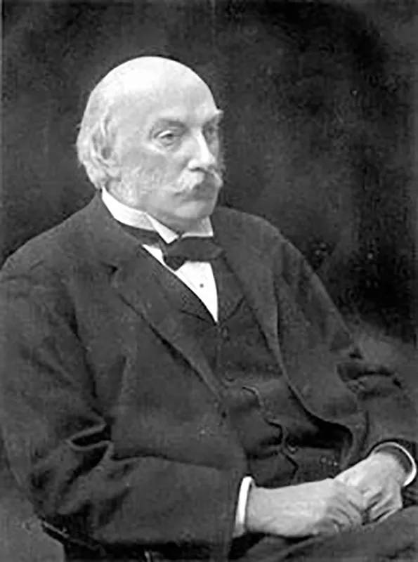
FIGURE 1.1 John William Strutt, 3rd Baron Rayleigh (1842–1919).
An understanding of the historical milestones of ultrasound require knowledge of transmission and pulse reflection methods, as well as A, B, and M modes of ultrasound. Early ultrasound used the transmission method where ultrasound measured the ultrasonic waves that passed through a specimen (6). The amount of sound waves not absorbed from the intervening tissue was recorded. The pulse reflection method placed the receiver and transmitter on the same side of the specimen and the amount of sound reflected was then recorded. Amplitude or A-mode was a one-dimensional image that displayed the amplitude or strength of a wave along the vertical axis and time on the horizontal axis (2). Brightness or B-mode, which is commonly used today, is a two-dimensional characterization of tissue where each dot or pixel on the screen represents an individual amplitude spike. In currently used models, amplitudes of varying intensity are assigned shades from black to white. M-motion or motion mode ultrasound relates the amplitude of the ultrasound wave to the imaging of moving structures (such as cardiac muscle).
DEVELOPMENT OF MEDICAL ULTRASONOGRAPHY
George Ludwig and Francis Struthers, working at the Naval Medical Research Institute in Bethesda, Maryland, were among the first to use echo technique in biologic tissue. Because they were employed by the military their work was considered restricted information and was not published in medical journals (2).
John Julian Wild was an English-trained surgeon who immigrated to the United States after World War II (Figure 1.2). Working in Owen Wangensteen’s laboratory at the University of Minnesota, using A-mode imaging and a 15 MHz transducer, Wild measured the thickness of the bowel wall in a large water tank (7). Ahead of his time, Wild felt that “it should be possible to detect tumors of the accessible portions of the gastroenterological tract both by density changes and also in all probability of the tumor tissue to fail to contract” (2). Wild managed to develop a scanning device that was used to screen patients for breast cancer and also developed transrectal and transvaginal transducers (2,8). With this instrument he imaged a brain tumor in a pathology specimen and localized a brain tumor in a patient after a craniotomy (2).
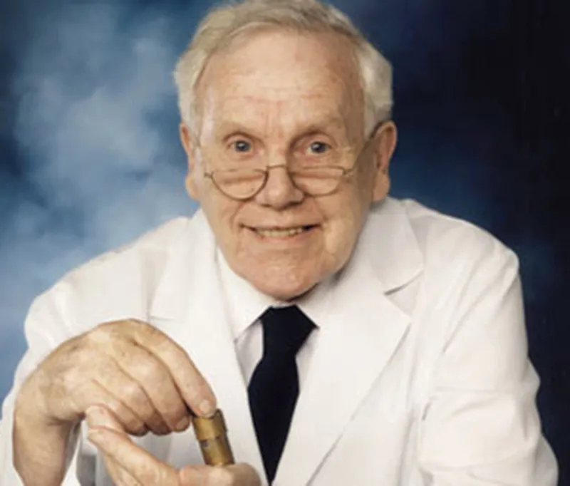
FIGURE 1.2 John Julian Wild (1914–2009).
Douglass Howry played an important role in the development of ultrasound and ultrasonic devices in the 1940s. Unlike Wild, Howry worked more on the development of the equipment and the applied theory of ultrasound rather than on its clinical application. Howry’s goal was to produce an instrument that was “in a manner comparable to the actual gross sectioning of structures in the pathological laboratory” (9). Working with W. Roderic Bliss, an electrical engineer, Howry built the first B-mode scanner in 1949. In the late 1950s, Howry and colleagues developed an ultrasound scanner with a semicircular pan containing a plastic window that was later developed into a direct-contact scanner. In 1961, Ralph Meyerdirk and William Wright produced the prototype for the first handheld contact scanner in the United States (10). Ian Donald at the University of Glasgow used an A-mode ultrasound machine to differentiate various types of tissues in recently excised fibroids and ovarian cysts. Donald and his colleagues in Glasgow contributed significant research in regard to ultrasonography in the field of obstetrics and gynecology. He incidentally discovered that a full urinary bladder provided a natural acoustic window for the transmission of ultrasound waves through the pelvis, thus allowing pelvic structures to be imaged more clearly (2,11).
TRANSRECTAL ULTRASONOGRAPHY
Anatomic ultrasound imaging is the oldest and most widely used technique to image the prostate. Watanabe et al. were the first to describe the use of transrectal ultrasound (TRUS) imaging of the prostate with a 3.5 MHz transducer in 1974 (12,13). Prostate cancer lesions have been classically described as hypoechoic lesions on TRUS imaging (14,15). However, the low specificity and positive predictive value (PPV) of TRUS imaging was a clear limitation as other conditions such as prostatitis or focal infarcts could also show hypoechoic characteristics. Lee et al. showed that the positive predictive value of TRUS by itself was 41%, the PPV dropped to 24% if the DRE was normal, 12% if the PSA was normal, and only 5% if both the DRE and PSA levels were normal (16). In 1989, Hodge et al. explored the utility of adding lesion-directed biopsies to specific hypoechoic areas found on ultrasound compared to the standard non-targeted TRUS-guided systematic prostate biopsy. In 136 men with abnormal prostates on DRE, they found that directed biopsies toward hypoechoic areas within the prostate added very little yield since in 80/83 patients, prostate cancer was detected by the non-targeted systematic biopsies. The addition of TRUS-guided cores to hypoechoic lesions increased the yield by only 5% (17). Thus TRUS imaging by itself has been inadequate for the diagnosis, characterization, and targeting of prostate cancer due to poor resolution, low specificity, and its negative predictive value. The main concerns of the technique include failure to detect prostate cancer (due to poor spatial resolution of lesions), inaccurate risk stratification (from under sampling in cases of small volume disease, transition zone or anteriorly located tumors) and high detection rate of small clinically insignificant prostate cancer (18,19,20). Most prostate cancer tissue is known to be harder or stiffer than normal prostate tissue (the digital rectal exam is predicated on the physician detecting harder or abnormal lumps of cancer tissue). With real-time elastography imaging tissue, the physician induces a mechanical excitation in the prostate tissue and then images the response using real-time ultrasound (21). Techniques include strain elastography, acoustic radiation force impulse imaging, and shear wave elastography. Despite some promising results, an absolute quantitative threshold to distinguish benign from malignant tissue still remains to be determined. Thus transrectal ultrasound guidance continues to be used principally to guide systematic 12-core biopsies of the prostate.
HISTORY OF MAGNETIC RESONANCE IMAGING
Nikola Tesla discovered the rotating magnetic field in 1882. In 1937, Columbia University Professor Isidor Rabi, working at the Pupin Physics Laboratory in New York City, recognized that the atomic nuclei show their presence by absorbing or emitting radio waves whe...