Gregory W.J. Hawryluk1, 2, Crystal A. Ruff1 and Michael G. Fehlings1, 2, 3*
1Division of Genetics and Development, Toronto Western Research Institute, Institute of Medical Science, University of Toronto, Toronto, Canada
2Division of Neurosurgery, University of Toronto, Toronto, Canada
3Spinal Program, University Health Network, Toronto Western Hospital, Toronto, Canada
*Correspondence to: Michael G. Fehlings MD, PhD, FRCSC, FACS, Krembil Chair, Neural Repair and Regeneration, Head, Spinal Program, University Health Network, Toronto Western Hospital, McLaughlin Pavilion, 12th floor Rm. 407, 399 Bathurst Street, Toronto, Ontario, Canada M5T 2S8. Tel: + 1-416-603-5627, Fax: + 1-416-603-5298, [email protected] The human central nervous system (CNS) may be the most complex structure in the universe. Its development and appropriate specification into phenotypically and spatially distinct neural subpopulations involves a precisely orchestrated response, with thousands of transcriptional regulators combining with epigenetic controls and specific temporal cues in perfect synchrony. Understandably, our insight into the sophisticated molecular mechanisms which underlie spinal cord development are as yet limited. Even less is known about abnormalities of this process – putative genetic and molecular causes of well-described defects have only begun to emerge in recent years. Nonetheless, modern scientific techniques are beginning to demonstrate common patterns and principles amid the tremendous complexity of spinal cord development and maldevelopment. These advances are important, given that developmental anomalies of the spinal cord are an important cause of mortality and morbidity (Sadler, 2000); it is hoped that research advances will lead to better methods to detect, treat, and prevent these lesions.
Gross embryology
Overview
A human term pregnancy lasts approximately 40 weeks, and the most dramatic and complex developmental processes are completed in the embryonic period spanning the first 8 weeks. In the embryonic period, critical developmental milestones include establishment of the midline and anteroposterior axis, formation of the three germ layers through gastrulation, and organogenesis. The subsequent fetal period is comparatively simple, wherein the developing human predominantly grows in size.
Fertilization to gastrulation
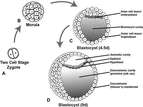
Fertilization characteristically takes place in the ampullary region of the fallopian tube. The fertilized egg then undergoes a number of mitotic divisions, eventually forming a 16-cell morula 3 days after fertilization (Fig. 1.1). Around the time the morula enters the uterus, it becomes known as a blastocyst and develops a cystic cavity known as a blastocele. By this time, the inner and outer cell masses have formed, which give rise to the embryo proper and the placenta respectively. The outer cell mass, also known as the trophoblast, secretes proteolytic enzymes which facilitate implantation in the endometrium, which occurs about 1 week following fertilization.
Fig. 1.1 Development of the bilaminar disc. The two-cell stage (A) is reached approximately 30 h after fertilization and the zygote eventually forms a 16-cell morula 2 days later (B). Inner and outer cell masses form at this time during a process referred to as compaction. The inner cell mass goes on to form the embryo, while the outer cell mass or trophoblast forms the placenta and extra-embryonic membranes. These masses become more apparent when the morula becomes a blastocyst 4.5 days after fertilization (C) and the blastocyst cavity develops. With further development the inner cell mass is known as the embryoblast. The blastocyst typically implants in the uterine mucosa 5–6 days after fertilization. The bilaminar disc forms within the blastocyst during the second week of development when the amniotic cavity develops within the epiblast (D). The constituent layers of the bilaminar disc are the epiblast (primitive ectoderm, lining the amniotic cavity) and the hypoblast (primitive endoderm, lining the primitive yolk sac).
In the second week of development, the inner cell mass, now known as the embryoblast, separates into two distinct cell layers, the hypoblast and the epiblast, which form the endoderm and ectoderm respectively. A second cystic cavity then develops adjacent to the epiblast. These layers thus form a bilaminar disc sandwiched between two cavities; the hypoblast lines the blastocyst cavity (primitive yolk sac) while the epiblast lines the developing amniotic cavity.
Gastrulation and Hensen’s node
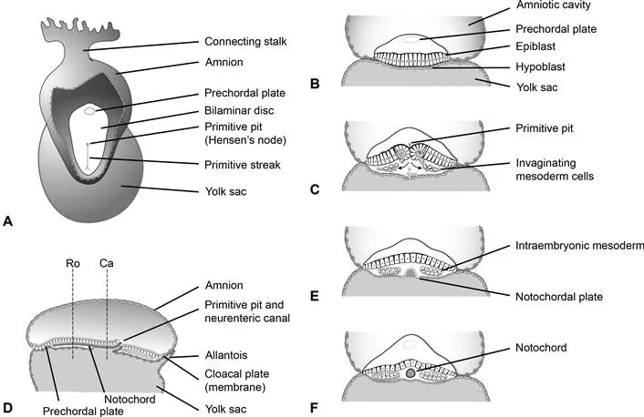
In the second week gastrulation occurs, which establishes the third germ layer, mesoderm (Fig. 1.2). Gastrulation begins with formation of the primitive streak in the caudal region of the epiblast. The cranial end of the primitive streak forms a thickening known variously as the primitive knot, the primitive node, or Hensen’s node. The primitive pit forms immediately posterior to the node and cells from the epiblast migrate here, invaginate, and then form intraembryonic endoderm and mesoderm.
Fig. 1.2 Gastrulation and development of the notochord. At the end of the second week of development a thickening of cells forms in the caudal midline of the bilaminar germ disc, referred to as the primitive streak (A). The prechordal plate is visible at the rostral end of the disc and eventually develops into the buccopharyngeal membrane. (B) and (C) show coronal views through the bilaminar disc. Epiblast cells invaginate at the primitive pit and primitive streak creating the cells of the definitive endoderm as well as the mesoderm through the process of gastrulation. Prenotochordal cells invaginate during this process and migrate as far rostral as the prechordal plate. Initially they intercalate with the hypoblast forming the notochordal plate (E). The notochordal plate then detaches from the endoderm, and forms a tube referred to as the definitive notochord (F). (E) and (F) are coronal views looking rostral from planes Ro and Ca shown in (D), which is a mid-sagittal section through the embryo at 17d postfertilization. The neurenteric canal is a temporary communication between the amniotic cavity and yolk sac believed to play a central role in many malformations of the spine and spinal cord.
The primitive node migrates caudally as gastrulation progresses, and although it typically regresses and forms the caudal eminence or end bud after migration to the sacrococcygeal area, it is deserving of some further discussion. Hensen’s node secretes morphogens such as fibroblast growth factor (FGF), sonic hedgehog (Shh) and retinoic acid (RA), playing key roles in neural induction and patterning which will be discussed in detail. In this fashion, Hensen’s node establishes the longitudinal axis, polarity and right–left sidedness within the embryo. It also participates in rostrocaudal specification along with paraxial mesoderm. Failure of Hensen’s node to regress can lead to formation of a sacrococcygeal teratoma.
Formation of the notochord
Another critical event occurring in the second week is the formation of the notochord (see Fig. 1.2). The notochord is a cylindrical structure derived from mesodermal cells which specifies the midline of the embryo, in addition to forming a rigid axis around which the embryo can develop. It also secretes inductive signals critical to the formation of the nervous system from the overlying ectoderm.
Prenotochordal cells which form the notochord migrate in from the primitive streak, and move rostrally toward the prechordal plate (future buccopharyngeal membrane) to form the notochordal process, a precursor of the notochord. The notochordal process initially intercalates with the hypoblast to form the notochordal plate. At this time an important transitory communication between the amniotic cavity and yolk sac forms which is known as the neurenteric canal. This canal is of great significance to spine and spinal cord maldevelopment, as it is currently believed to play a critical role in numerous malformations such as neurenteric cysts and split cord malformations (Pang and Dias, 1992), as will be discussed.
Almost immediately after the notochordal plate forms and directly contacts the yolk sac, it separates from the endoderm, moves slightly dorsally and re-forms a cord of cells running along the rostrocaudal axis of the embryo’s midline. Despite its embryological significance, few notochord remnants persist in the adult. These cells make up the nucleus pulposus at the center of the intervertebral disc and notochord remnants are also believed to give rise to chordomas and notochordal rests (Kyriakos et al., 2003).
Primary neurulation
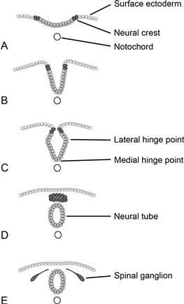
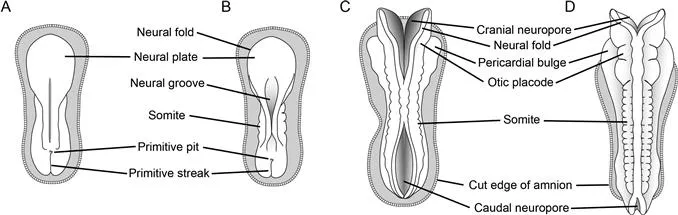
The central nervous system begins to develop in the third week postfertilization and the process begins with neurulation (Figs 1.3, 1.4). At the outset of neurulation, the notochord induces a subset of ectodermal cells to differentiate into neural precursor cells, forming a columnar epithelium referred to as the neural plate. Primary neurulation occurs when the neural plate folds and closes to form the neural tube.
Fig. 1.3 Primary neurulation. The neural tube and cells of the neural crest are derived from surface ectoderm, forming a columnar epithelium referred to as the neural plate, as a result of induction by the notochord. Neural crest cells initially reside lateral to those that will form the neural tube (A). Folding internalizes these cells (B). Medial and lateral hinge points serve to anchor the neural tube, facilitating this folding (C). The medial hinge point is also known as the floor plate. The neural crest cells separate and form a mass dorsal to the neural tube (D). They later migrate to form dorsal root ganglia and many important cells types within the embryo (E).
Fig. 1.4 Closure of the neural tube. Dorsal views of the embryo are shown with the amnion removed. (A) An 18-day-old embryo with a prominent neural plate is about to undergo primary neurulation. (B) At 20 days post‐fertilization, somites begin to appear and the neural folds begin to meet and fuse; this proc...