The Unofficial Guide to Radiology
Chest, Abdominal, Orthopaedic X Rays, plus CTs, MRIs and Other Important Modalities
Mark Rodrigues, Zeshan Qureshi
- 850 pagine
- English
- ePUB (disponibile sull'app)
- Disponibile su iOS e Android
The Unofficial Guide to Radiology
Chest, Abdominal, Orthopaedic X Rays, plus CTs, MRIs and Other Important Modalities
Mark Rodrigues, Zeshan Qureshi
Informazioni sul libro
X-ray interpretation is an important part of clinical work for all doctors. Unfortunately it is often an overlooked subject in the medical school curriculum, which many medical students and junior doctors find difficult and daunting. From the same series as The Unofficial Guide to Passing OSCEs, The Unofficial Guide to Radiology aims to remedy this by providing a systematic approach to chest, abdominal and musculoskeletal X-ray interpretation. It is designed to be a useful learning resource for medical students, junior and hospital doctors, nurse practitioners and radiology trainees. The chest, abdominal and musculoskeletal X-ray chapters contain step-by-step approaches to interpreting and presenting X-rays. Each of these chapters then covers 20 common and important X-ray cases/diagnoses, which a junior doctor should be able to confidently identify. The content is in line with the Royal College of Radiologists' Undergraduate Radiology Curriculum 2012, making it up to date and relevant to today's students and junior doctors. The layout is designed to make the book as clinically relevant as possible; the X-rays are presented in the context of a clinical scenario. The reader is asked to "present their findings" before turning over the page to reveal a model X-ray report accompanied by a fully annotated version of the X-ray. This encourages the reader to look at the X-ray thoroughly, as if working on a ward, and come to their own conclusions before seeing the answers. To further enhance the clinical relevance, each case has 5 clinical and radiology-related multiple-choice questions with detailed answers. These are aimed to test core knowledge needed for exams and working life, and illustrate how the X-ray findings will influence patient management. One of the keys to X-ray interpretation is practice, practice and more practice. The bonus X-ray chapter provides over 50 further X - ray cases to help consolidate the reader's knowledge and provide an opportunity to practice the skills they have learnt. In addition to these four core chapters the introductory chapter covers the (very) basic science behind X-rays, the relevant legislation controlling X-rays and tips on how to request radiology examinations. Additionally a chapter is devoted to other important imaging investigations, such as computed tomography (CT), magnetic resonance imaging (MRI) and ultrasound, covering the details of what the examinations involve, their common indications and contraindications and key imaging findings. The Unofficial Guide to Radiology is written by both radiologists and clinicians, and reviewed by a panel of medical students to ensure its relevance.
Domande frequenti
Informazioni
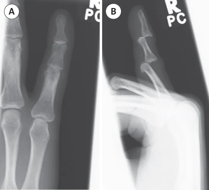
ORTHOPAEDIC X-RAYS
| Spine Case 1 | Wrist Case 1 | Hip Case 3 | Knee Case 2 |
| Spine Case 2 | Wrist Case 2 | Hip Case 4 | Knee Case 3 |
| Spine Case 3 | Wrist Case 3 | Hip Case 5 | Tibia/Fibula Case 1 |
| Shoulder Case 1 | Hip Case 1 | Hip Case 6 | Tibia/Fibula Case 2 |
| Elbow Case 1 | Hip Case 2 | Knee Case 1 | Ankle Case 1 |
| Systematic approach to orthopaedic X-rays 1.Projection 2.Patient details 3.Technical adequacy 4.Obvious abnormalities 5.Systematic review of the X-ray 6.Summary |
1. Projection
| Remember: children develop and mature at different rates and therefore the age of a patient does not tell you whether they are skeletally mature or not. |