The Unofficial Guide to Radiology
eBook - ePub

The Unofficial Guide to Radiology

Chest, Abdominal, Orthopaedic X Rays, plus CTs, MRIs and Other Important Modalities

Mark Rodrigues, Zeshan Qureshi

Partager le livre
  1. 850 pages
  2. English
  3. ePUB (adapté aux mobiles)
  4. Disponible sur iOS et Android
eBook - ePub

The Unofficial Guide to Radiology

Chest, Abdominal, Orthopaedic X Rays, plus CTs, MRIs and Other Important Modalities

Mark Rodrigues, Zeshan Qureshi

DĂ©tails du livre
Aperçu du livre
Table des matiĂšres
Citations

À propos de ce livre

X-ray interpretation is an important part of clinical work for all doctors. Unfortunately it is often an overlooked subject in the medical school curriculum, which many medical students and junior doctors find difficult and daunting. From the same series as The Unofficial Guide to Passing OSCEs, The Unofficial Guide to Radiology aims to remedy this by providing a systematic approach to chest, abdominal and musculoskeletal X-ray interpretation. It is designed to be a useful learning resource for medical students, junior and hospital doctors, nurse practitioners and radiology trainees. The chest, abdominal and musculoskeletal X-ray chapters contain step-by-step approaches to interpreting and presenting X-rays. Each of these chapters then covers 20 common and important X-ray cases/diagnoses, which a junior doctor should be able to confidently identify. The content is in line with the Royal College of Radiologists' Undergraduate Radiology Curriculum 2012, making it up to date and relevant to today's students and junior doctors. The layout is designed to make the book as clinically relevant as possible; the X-rays are presented in the context of a clinical scenario. The reader is asked to "present their findings" before turning over the page to reveal a model X-ray report accompanied by a fully annotated version of the X-ray. This encourages the reader to look at the X-ray thoroughly, as if working on a ward, and come to their own conclusions before seeing the answers. To further enhance the clinical relevance, each case has 5 clinical and radiology-related multiple-choice questions with detailed answers. These are aimed to test core knowledge needed for exams and working life, and illustrate how the X-ray findings will influence patient management. One of the keys to X-ray interpretation is practice, practice and more practice. The bonus X-ray chapter provides over 50 further X - ray cases to help consolidate the reader's knowledge and provide an opportunity to practice the skills they have learnt. In addition to these four core chapters the introductory chapter covers the (very) basic science behind X-rays, the relevant legislation controlling X-rays and tips on how to request radiology examinations. Additionally a chapter is devoted to other important imaging investigations, such as computed tomography (CT), magnetic resonance imaging (MRI) and ultrasound, covering the details of what the examinations involve, their common indications and contraindications and key imaging findings. The Unofficial Guide to Radiology is written by both radiologists and clinicians, and reviewed by a panel of medical students to ensure its relevance.

Foire aux questions

Comment puis-je résilier mon abonnement ?
Il vous suffit de vous rendre dans la section compte dans paramĂštres et de cliquer sur « RĂ©silier l’abonnement ». C’est aussi simple que cela ! Une fois que vous aurez rĂ©siliĂ© votre abonnement, il restera actif pour le reste de la pĂ©riode pour laquelle vous avez payĂ©. DĂ©couvrez-en plus ici.
Puis-je / comment puis-je télécharger des livres ?
Pour le moment, tous nos livres en format ePub adaptĂ©s aux mobiles peuvent ĂȘtre tĂ©lĂ©chargĂ©s via l’application. La plupart de nos PDF sont Ă©galement disponibles en tĂ©lĂ©chargement et les autres seront tĂ©lĂ©chargeables trĂšs prochainement. DĂ©couvrez-en plus ici.
Quelle est la différence entre les formules tarifaires ?
Les deux abonnements vous donnent un accĂšs complet Ă  la bibliothĂšque et Ă  toutes les fonctionnalitĂ©s de Perlego. Les seules diffĂ©rences sont les tarifs ainsi que la pĂ©riode d’abonnement : avec l’abonnement annuel, vous Ă©conomiserez environ 30 % par rapport Ă  12 mois d’abonnement mensuel.
Qu’est-ce que Perlego ?
Nous sommes un service d’abonnement Ă  des ouvrages universitaires en ligne, oĂč vous pouvez accĂ©der Ă  toute une bibliothĂšque pour un prix infĂ©rieur Ă  celui d’un seul livre par mois. Avec plus d’un million de livres sur plus de 1 000 sujets, nous avons ce qu’il vous faut ! DĂ©couvrez-en plus ici.
Prenez-vous en charge la synthÚse vocale ?
Recherchez le symbole Écouter sur votre prochain livre pour voir si vous pouvez l’écouter. L’outil Écouter lit le texte Ă  haute voix pour vous, en surlignant le passage qui est en cours de lecture. Vous pouvez le mettre sur pause, l’accĂ©lĂ©rer ou le ralentir. DĂ©couvrez-en plus ici.
Est-ce que The Unofficial Guide to Radiology est un PDF/ePUB en ligne ?
Oui, vous pouvez accĂ©der Ă  The Unofficial Guide to Radiology par Mark Rodrigues, Zeshan Qureshi en format PDF et/ou ePUB ainsi qu’à d’autres livres populaires dans Medicina et Teoria, pratica e riferimenti medici. Nous disposons de plus d’un million d’ouvrages Ă  dĂ©couvrir dans notre catalogue.

Informations

Année
2014
ISBN
9781910399071

ORTHOPAEDIC X-RAYS

Spine Case 1 Wrist Case 1 Hip Case 3 Knee Case 2
Spine Case 2 Wrist Case 2 Hip Case 4 Knee Case 3
Spine Case 3 Wrist Case 3 Hip Case 5 Tibia/Fibula Case 1
Shoulder Case 1 Hip Case 1 Hip Case 6 Tibia/Fibula Case 2
Elbow Case 1 Hip Case 2 Knee Case 1 Ankle Case 1
This introduction to the chapter is aimed at providing a systematic framework for approaching orthopaedic X-rays. Further details and examples of the specific X-rays findings discussed below are covered more extensively in the example cases later in the chapter and the bonus X-ray chapter.
KEY POINT
Systematic approach to orthopaedic X-rays
1.Projection
2.Patient details
3.Technical adequacy
4.Obvious abnormalities
5.Systematic review of the X-ray
6.Summary

1. Projection

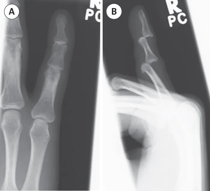
‱Assessment of any bone or joint in general requires at least two views–‘one view is one too few!’ These normally consist of AP and lateral X-rays (figure 1).
‱For some sites, such as the scaphoid, where fractures are difficult to detect, it is routine to obtain more than two views.
‱For some patients in whom the clinical suspicion of a fracture is high but is not evident on the usual two views, additional views may be requested, such as internal rotation views of the hip.
‱If the shaft of a long bone is fractured it is imperative to X-ray the joint above and below because of the potential for additional injuries (fracture or dislocation) at these sites.
‱Comment on whether the patient is skeletally mature (fused epiphyses/growth plates). This is useful because the types of injury and pathology vary between skeletally immature and mature patients.
KEY POINT
Remember: children develop and mature at different rates and therefore the age of a patient does not tell you whether they are skeletally mature or not.
image
Figure 1.
Image A: AP view of this finger shows apparently normal alignment of the interphalangeal joints
Image B: The lateral view shows posterior dislocations at both of these joints!
These X-rays dramatically show why one view is one too few!

2. Patient Details

‱It is important to check you are looking at the correct X-ray from the correct patient.
‱The patient details should be listed on the X-ray.
‱State the name, age (or date of birth), and the date on which the X-ray was taken.

3. Technical Adequacy

‱The entire area of concern should be included in the X-ray. This is particularly important with X-rays of the cervical spine.
‱Are the X-rays adequately exposed so that bone and soft tissues can be seen and differentiated?
‱Is the patient rotated? Sometimes a small amount of rotation, which results in a nonstandard view, can make accurate assessment difficult. If the patient is significantly rotated, then the X-rays may need to be repeated.

4. Obvious Abnormalities

‱If there is an obvious abnormality, such as fractures, subluxations/dislocations, or bone lesions, comment on this before conducting your systemic review of the X-ray.

5. Systematic Review of the X-ray (Figures 2-5)

‱Look around the edges of all the bones for fractures. The cortex should be a smooth, continuous line. Any disruption to, or irregularity of this may represent a fracture.
‱Also look at the medulla for evidence of fractures – look for disruption to the trabeculations and for lucent and sclerotic lines.
‱Displaced fractures appear as black lines, whereas impacted or overlapping fractures usually result in sclerotic areas.
‱Assess for soft tissue swelling and joint effusion (knee and elbow joints), if appropriate. Such findings may represent soft tissue injury or can be indirect evidence of a fracture.
‱Look at the joint surfaces for any evidence of subluxation or dislocation.
‱Assess joints for degenerative (loss of joint space, subchondral sclerosis, subchondral cysts and osteophytes) and inflammatory changes (periarticular osteoporosis, soft tissue swelling and bony erosions).
‱Review the bon...

Table des matiĂšres