
eBook - ePub
Neuroradiology
Spectrum and Evolution of Disease
Juan Small, Daniel Noujaim, Daniel T. Thomas Ginat, Hillary R Kelly, Pamela W Schaefer
This is a test
Condividi libro
- 600 pagine
- English
- ePUB (disponibile sull'app)
- Disponibile su iOS e Android
eBook - ePub
Neuroradiology
Spectrum and Evolution of Disease
Juan Small, Daniel Noujaim, Daniel T. Thomas Ginat, Hillary R Kelly, Pamela W Schaefer
Dettagli del libro
Anteprima del libro
Indice dei contenuti
Citazioni
Informazioni sul libro
Acquire a better understanding of disease evolution and treatment response with Neuroradiology Spectrum and Evolution of Disease. The unique format includes carefully chosen clinical images that depict the pathologic evolution of disease from initial presentation across the continuum of progression. Colorful graphics plot characteristic changes, helping you visualize how normal and abnormal variations alter over time. Extensive illustrations and concise descriptions distill complex concepts, making this first-of-its-kind resource an excellent tool for imaging interpretation and clinical problem solving.
- Presents neurologic disease from a novel imaging perspective, emphasizing evolutionary development and pointing out patterns to recognize.
- Provides a state-of-the-art understanding of image interpretation based on early, middle, and late imaging characteristics; typical and atypical variants; and pre-treatment, post-treatment, progression, and regression characteristics.
- Guides you through the progression of disease with chronological indicators, additional clinical images and descriptions, and annotations that highlight atypical findings – for an easy-to-digest, visually memorable presentation.
- Helps you correctly interpret specific imaging characteristics you have never seen, even when the disease process is one you are familiar with.
Domande frequenti
Come faccio ad annullare l'abbonamento?
È semplicissimo: basta accedere alla sezione Account nelle Impostazioni e cliccare su "Annulla abbonamento". Dopo la cancellazione, l'abbonamento rimarrà attivo per il periodo rimanente già pagato. Per maggiori informazioni, clicca qui
È possibile scaricare libri? Se sì, come?
Al momento è possibile scaricare tramite l'app tutti i nostri libri ePub mobile-friendly. Anche la maggior parte dei nostri PDF è scaricabile e stiamo lavorando per rendere disponibile quanto prima il download di tutti gli altri file. Per maggiori informazioni, clicca qui
Che differenza c'è tra i piani?
Entrambi i piani ti danno accesso illimitato alla libreria e a tutte le funzionalità di Perlego. Le uniche differenze sono il prezzo e il periodo di abbonamento: con il piano annuale risparmierai circa il 30% rispetto a 12 rate con quello mensile.
Cos'è Perlego?
Perlego è un servizio di abbonamento a testi accademici, che ti permette di accedere a un'intera libreria online a un prezzo inferiore rispetto a quello che pagheresti per acquistare un singolo libro al mese. Con oltre 1 milione di testi suddivisi in più di 1.000 categorie, troverai sicuramente ciò che fa per te! Per maggiori informazioni, clicca qui.
Perlego supporta la sintesi vocale?
Cerca l'icona Sintesi vocale nel prossimo libro che leggerai per verificare se è possibile riprodurre l'audio. Questo strumento permette di leggere il testo a voce alta, evidenziandolo man mano che la lettura procede. Puoi aumentare o diminuire la velocità della sintesi vocale, oppure sospendere la riproduzione. Per maggiori informazioni, clicca qui.
Neuroradiology è disponibile online in formato PDF/ePub?
Sì, puoi accedere a Neuroradiology di Juan Small, Daniel Noujaim, Daniel T. Thomas Ginat, Hillary R Kelly, Pamela W Schaefer in formato PDF e/o ePub, così come ad altri libri molto apprezzati nelle sezioni relative a Medicine e Neurology. Scopri oltre 1 milione di libri disponibili nel nostro catalogo.
Section I
Brain
Parenchymal Hemorrhage and Trauma
1
Brain Parenchymal Hematoma Evolution
Juan E. Small
Key Words
Brain; Parenchymal; Hemorrhage; Hematoma
Introduction
Magnetic resonance imaging (MRI) can differentiate between acute, subacute, and chronic hemorrhage because of its sensitivity and specificity to hemoglobin degradation products. Therefore the imaging interpreter is, with proper knowledge, able to estimate the age of a brain parenchymal hematoma. The blood products in a hematoma evolve through a predictable variation in hemoglobin oxygenation states and hemoglobin byproducts. This predictable pattern of hematoma evolution over time leads to a specific pattern of changing signal intensities on conventional MRI.
There are limitations to the accuracy of hematoma age interpretation. Several direct and indirect factors, including the operating field strength of the magnet, the mode of image acquisition, and a wide range of biologic factors particular to the patient, may affect the imaging evolution of a parenchymal hematoma. Despite substantial variability, it is generally accepted that five stages of parenchymal hemorrhage can be distinguished by MRI. A basic understanding of the biochemical evolution of brain parenchymal hemorrhage and magnetic properties that affect MRI signal are essential for interpretation.
Temporal Evolution: Overview
A well-described pathophysiologic process of evolution and resorption for parenchymal hemorrhage involves five distinct phases (Fig. 1.1).
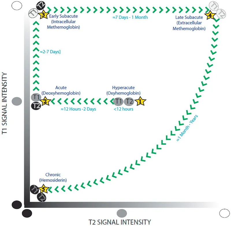
Figure 1.1 Five stages of parenchymal hematoma evolution on magnetic resonance imaging. One can easily remember the T1 and T2 characteristics of an evolving hematoma by memorizing this figure. Start from the center of the figure and move according to the direction of the arrows to remember the signal characteristics of the five distinct phases of hematoma evolution.
With this knowledge, the imaging interpreter can often identify the relative age of a brain parenchymal hematoma based on the T1 and T2 characteristics of the collection. However, it is important to realize that hematoma evolution is a fluid process (without static or punctuated steps). Therefore, stages of hemorrhage commonly coexist within the same hematoma because hemoglobin degradation proceeds at variable rates in the center versus the periphery of a single hematoma cavity. By convention, the most mature form of hemoglobin present defines the stage of hematoma evolution (Fig. 1.2).
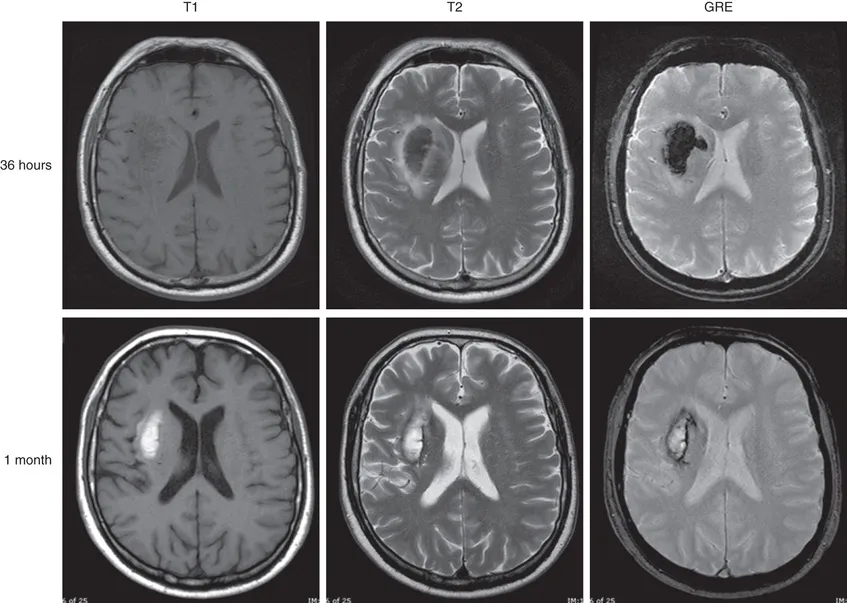
Figure 1.2 Parenchymal hemorrhage at 36 hours and at 1 month after initial presentation. At 36 hours (acute phase), T2 signal is low and T1 signal remains intermediate consistent with intracellular deoxyhemoglobin. There is diffuse hypointensity on the gradient echo (GRE) image due to the paramagnetic effects of deoxyhemoglobin. At 1 month (end of late subacute phase), central T1 and T2 hyperintensity is consistent with extracellular methemoglobin, while peripheral T1 and T2 low signal is consistent with hemosiderin. The rim is hypointense on the GRE image due to the superparamagnetic effects of hemosiderin. The hematoma is smaller due to retraction.
Temporal Evolution: in Greater Depth
Extravascular blood in a hemorrhagic collection remains as oxyhemoglobin for 2 to 3 hours. The immediate activation of the clotting cascade begins the process of clot formation. Deoxyhemoglobin begins to form at the periphery of the hematoma. Eventually, the failure of metabolic pathways preventing oxidation of heme iron results in conversion of hemoglobin to methemoglobin.
In the hyperacute stage, parenchymal hemorrhage is a liquid almost completely composed of intracellular oxygenated hemoglobin. Over the course of a few hours, a heterogeneous blood clot forms within the hematoma cavity, composed of red blood cells, platelets, and serum. In the acute phase, intracellular hemoglobin becomes deoxygenated. Vasogenic edema develops in the surrounding brain parenchyma. In the early subacute phase, deoxyhemoglobin is gradually converted to intracellular methemoglobin. Then, in the late subacute phase, lysis of red blood cells leads to the release of methemoglobin into the extracellular space. During this time, the surrounding vasogenic edema slowly begins to decrease and the clot slowly retracts. In the chronic stage, macrophages and glial cells phagocytose the hematoma, leading to intracellular ferritin and hemosiderin. Eventually, the hematoma resolves and leaves a posthemorrhagic cavity with hemosiderin-stained walls.
It is critical to realize that Fig. 1.1 represents a simplified version of events designed to aid in memory. As noted previously, hematoma evolution is a fluid process (without static or punctuated steps). Stages of hemorrhage commonly coexist within the same hematoma because hemoglobin degradation proceeds at variable rates in the center versus the periphery of a single hematoma cavity (Figs. 1.3 and 1.4).
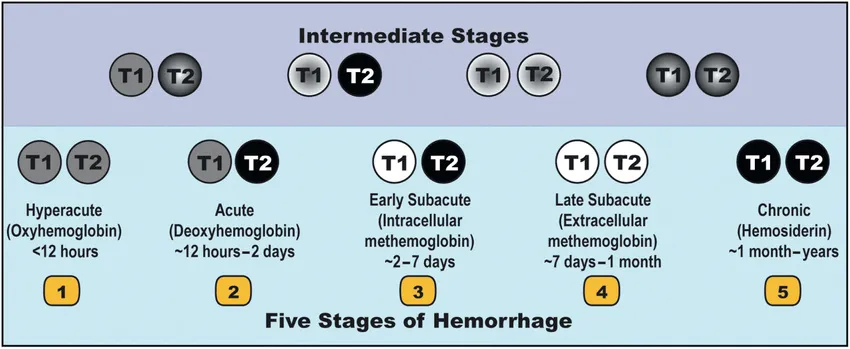
Figure 1.3 By convention, the most mature form of hemoglobin present defines the stage of hematoma evolution. The five stages of hemorrhage depicted in Fig. 1.1 are seen in the bottom row of Fig. 1.3. Each stage depicts the most mature form of hemoglobin present in the hematoma. They are not meant to imply homogeneity. The top row depicts intermediate stages of hematoma evolution between each step. In the intermediate stages, the most mature form of hemorrhage is seen at the periphery.
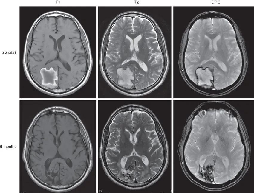
Figure 1.4 Parenchymal hemorrhage 25 days and 6 months after initial presentation. At 25 days (between early subacute to late subacute phase), there is ...