
eBook - ePub
Surgical anatomy of the lateral transpsoas approach to the lumbar spine E-Book
R. Shane Tubbs, Rod J. Oskouian, Jr., Joe Iwanaga, Marc Moisi
This is a test
Condividi libro
- 256 pagine
- English
- ePUB (disponibile sull'app)
- Disponibile su iOS e Android
eBook - ePub
Surgical anatomy of the lateral transpsoas approach to the lumbar spine E-Book
R. Shane Tubbs, Rod J. Oskouian, Jr., Joe Iwanaga, Marc Moisi
Dettagli del libro
Anteprima del libro
Indice dei contenuti
Citazioni
Informazioni sul libro
Surgical anatomy of the lateral transpsoas approach to the lumbar spine E-Book
Domande frequenti
Come faccio ad annullare l'abbonamento?
È semplicissimo: basta accedere alla sezione Account nelle Impostazioni e cliccare su "Annulla abbonamento". Dopo la cancellazione, l'abbonamento rimarrà attivo per il periodo rimanente già pagato. Per maggiori informazioni, clicca qui
È possibile scaricare libri? Se sì, come?
Al momento è possibile scaricare tramite l'app tutti i nostri libri ePub mobile-friendly. Anche la maggior parte dei nostri PDF è scaricabile e stiamo lavorando per rendere disponibile quanto prima il download di tutti gli altri file. Per maggiori informazioni, clicca qui
Che differenza c'è tra i piani?
Entrambi i piani ti danno accesso illimitato alla libreria e a tutte le funzionalità di Perlego. Le uniche differenze sono il prezzo e il periodo di abbonamento: con il piano annuale risparmierai circa il 30% rispetto a 12 rate con quello mensile.
Cos'è Perlego?
Perlego è un servizio di abbonamento a testi accademici, che ti permette di accedere a un'intera libreria online a un prezzo inferiore rispetto a quello che pagheresti per acquistare un singolo libro al mese. Con oltre 1 milione di testi suddivisi in più di 1.000 categorie, troverai sicuramente ciò che fa per te! Per maggiori informazioni, clicca qui.
Perlego supporta la sintesi vocale?
Cerca l'icona Sintesi vocale nel prossimo libro che leggerai per verificare se è possibile riprodurre l'audio. Questo strumento permette di leggere il testo a voce alta, evidenziandolo man mano che la lettura procede. Puoi aumentare o diminuire la velocità della sintesi vocale, oppure sospendere la riproduzione. Per maggiori informazioni, clicca qui.
Surgical anatomy of the lateral transpsoas approach to the lumbar spine E-Book è disponibile online in formato PDF/ePub?
Sì, puoi accedere a Surgical anatomy of the lateral transpsoas approach to the lumbar spine E-Book di R. Shane Tubbs, Rod J. Oskouian, Jr., Joe Iwanaga, Marc Moisi in formato PDF e/o ePub, così come ad altri libri molto apprezzati nelle sezioni relative a Medicine e Neurosurgery. Scopri oltre 1 milione di libri disponibili nel nostro catalogo.
Informazioni
Argomento
MedicineCategoria
NeurosurgeryChapter 1
Superficial Nerves of the Anterolateral Abdominal Wall and the Lateral Transpsoas Approach to the Lumbar Spine
Mary Katherine Cleveland, Joe Iwanaga, and R. Shane Tubbs
Abstract
Lateral approaches to the lumbar spine have increased in popularity and offer an additional option to surgeons. However, these approaches demand a good working knowledge of regional anatomy in order to avoid complications. Herein, we describe the anatomy of the superficial nerves related to this approach.
Keywords
Anatomy; Lumbar; Spine; Surgery; Complications
Introduction
The lateral transpsoas approach to the lumbar spine is increasingly being used to treat degenerative changes requiring fusion (Arnold et al., 2012). In contrast to conventional posterior spinal fusion techniques, this minimally invasive approach spares extensive posterior tissue dissection and resection and decreases operative time, blood loss, postoperative pain, and tissue trauma. Although minimally invasive, this procedure has the approach-related risk to cause lumbar plexus nerve injuries secondary to the insertion and dilation of dilatators or retractors. Plexus injuries are reported in up to approximately 30% of patients, often presenting as neuropathic pain and motor or sensory deficits (Arnold et al., 2012; Pumberger et al., 2012; Rodgers et al., 2011).
Several anatomical studies on plexus nerve anatomy for lateral approaches have been published (Uribe et al., 2010; Benglis et al., 2009); however, few have systematically documented the types of injury typically observed at each spinal level after such procedures. In our earlier study, approximately 50% of all operated-upon segments had plexus nerve injuries occurring at segments L1–L4 and involving nerve roots as well as motor and sensory nerves (Grunert et al., 2017).
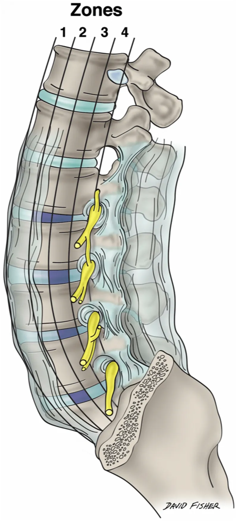
Moro and Uribe et al. subdivided each lumbar vertebral segment into four quarters (zone I to zone IV) from the anterior to the posterior border of the vertebral body (Fig. 1.1) (Uribe et al., 2010; Moro et al., 2003). Although describing the relationship of the plexus nerves to the lateral vertebral body surface helps in determining ideal docking points, it oversimplifies the complex plexus anatomy, as the nerves run in anteroposterior, superolateral, and mediolateral directions. As shown in the study of Grunert et al., injuries can occur throughout the entire trajectory of the lateral transpsoas approach to the lumbar spine. Over 50% of the nerve injuries occurred either at the lateral aspect of the psoas major muscle, within the outer abdominal muscles, or in the subcutaneous tissue of the abdominal wall, predominantly affecting the subcostal, ilioinguinal, iliohypogastric, and lateral femoral cutaneous nerves (Grunert et al., 2017).
As the superficial nerves of the region between the iliac crest and 12th rib are so concentrated (Fig. 1.2) and important to avoid with the lateral transpsoas approach to the lumbar spine, this chapter focuses on these structures and their detailed anatomy.
Segmental Lateral Cutaneous Branches
The skin and muscles of the anterolateral abdominal wall are innervated by the ventral rami of T7–T12. The muscles here also receive fibers from at least L1 ventral ramus and, as we have reported, as inferior as an L4 contribution. The lower thoracic and L1 nerves and their branches travel into the abdominal wall between the transversus abdominis and internal abdominal oblique muscles. Distally, they pierce the rectus sheath. Along their course, the nerves supply not only the skin and adjacent musculature but also the parietal peritoneum. These nerves give rise to lateral and anterior cutaneous branches (Figs. 1.3 and 1.4). The former arise at about the anterior axillary line and pierce the anterolateral muscles of the abdominal wall near the midaxillary line. As the lateral cutaneous branches of these nerves reach the skin, they split into anterior and posterior branches. The anterior cutaneous branches are the terminal branches of each of these segmental nerves and exit the rectus sheath anteriorly to reach the overlying skin where they too split into branches, medial and lateral.
Superior Cluneal Nerves
The superior cluneal nerves (SCNs) (Figs. 1.5–1.8) are the posterior cutaneous branches (from the lateral branch) of the dorsal rami usually described as arising from the upper three lumbar spinal nerves. Historically, it has been believed that the origin of the SCN is the dorsal rami of the L1, L2, and L3 spinal nerves. The SCN is usually depicted as having three branches: medial, intermediate, and lateral SCN.
Out of 20 sides, we previously reported the vertebral level of the origin of the SCN was T12 on 2 sides (10%), L1 on 15 sides (75%), L2 on 18 sides (90%), L3 on 19 sides (95%), L4 on 9 sides (45%) (Fig. 1.9), and L5 on 2 sides (10%), respec...