
eBook - ePub
Surgical anatomy of the lateral transpsoas approach to the lumbar spine E-Book
R. Shane Tubbs, Rod J. Oskouian, Jr., Joe Iwanaga, Marc Moisi
This is a test
Buch teilen
- 256 Seiten
- English
- ePUB (handyfreundlich)
- Über iOS und Android verfügbar
eBook - ePub
Surgical anatomy of the lateral transpsoas approach to the lumbar spine E-Book
R. Shane Tubbs, Rod J. Oskouian, Jr., Joe Iwanaga, Marc Moisi
Angaben zum Buch
Buchvorschau
Inhaltsverzeichnis
Quellenangaben
Über dieses Buch
Surgical anatomy of the lateral transpsoas approach to the lumbar spine E-Book
Häufig gestellte Fragen
Wie kann ich mein Abo kündigen?
Gehe einfach zum Kontobereich in den Einstellungen und klicke auf „Abo kündigen“ – ganz einfach. Nachdem du gekündigt hast, bleibt deine Mitgliedschaft für den verbleibenden Abozeitraum, den du bereits bezahlt hast, aktiv. Mehr Informationen hier.
(Wie) Kann ich Bücher herunterladen?
Derzeit stehen all unsere auf Mobilgeräte reagierenden ePub-Bücher zum Download über die App zur Verfügung. Die meisten unserer PDFs stehen ebenfalls zum Download bereit; wir arbeiten daran, auch die übrigen PDFs zum Download anzubieten, bei denen dies aktuell noch nicht möglich ist. Weitere Informationen hier.
Welcher Unterschied besteht bei den Preisen zwischen den Aboplänen?
Mit beiden Aboplänen erhältst du vollen Zugang zur Bibliothek und allen Funktionen von Perlego. Die einzigen Unterschiede bestehen im Preis und dem Abozeitraum: Mit dem Jahresabo sparst du auf 12 Monate gerechnet im Vergleich zum Monatsabo rund 30 %.
Was ist Perlego?
Wir sind ein Online-Abodienst für Lehrbücher, bei dem du für weniger als den Preis eines einzelnen Buches pro Monat Zugang zu einer ganzen Online-Bibliothek erhältst. Mit über 1 Million Büchern zu über 1.000 verschiedenen Themen haben wir bestimmt alles, was du brauchst! Weitere Informationen hier.
Unterstützt Perlego Text-zu-Sprache?
Achte auf das Symbol zum Vorlesen in deinem nächsten Buch, um zu sehen, ob du es dir auch anhören kannst. Bei diesem Tool wird dir Text laut vorgelesen, wobei der Text beim Vorlesen auch grafisch hervorgehoben wird. Du kannst das Vorlesen jederzeit anhalten, beschleunigen und verlangsamen. Weitere Informationen hier.
Ist Surgical anatomy of the lateral transpsoas approach to the lumbar spine E-Book als Online-PDF/ePub verfügbar?
Ja, du hast Zugang zu Surgical anatomy of the lateral transpsoas approach to the lumbar spine E-Book von R. Shane Tubbs, Rod J. Oskouian, Jr., Joe Iwanaga, Marc Moisi im PDF- und/oder ePub-Format sowie zu anderen beliebten Büchern aus Medicine & Neurosurgery. Aus unserem Katalog stehen dir über 1 Million Bücher zur Verfügung.
Information
Thema
MedicineThema
NeurosurgeryChapter 1
Superficial Nerves of the Anterolateral Abdominal Wall and the Lateral Transpsoas Approach to the Lumbar Spine
Mary Katherine Cleveland, Joe Iwanaga, and R. Shane Tubbs
Abstract
Lateral approaches to the lumbar spine have increased in popularity and offer an additional option to surgeons. However, these approaches demand a good working knowledge of regional anatomy in order to avoid complications. Herein, we describe the anatomy of the superficial nerves related to this approach.
Keywords
Anatomy; Lumbar; Spine; Surgery; Complications
Introduction
The lateral transpsoas approach to the lumbar spine is increasingly being used to treat degenerative changes requiring fusion (Arnold et al., 2012). In contrast to conventional posterior spinal fusion techniques, this minimally invasive approach spares extensive posterior tissue dissection and resection and decreases operative time, blood loss, postoperative pain, and tissue trauma. Although minimally invasive, this procedure has the approach-related risk to cause lumbar plexus nerve injuries secondary to the insertion and dilation of dilatators or retractors. Plexus injuries are reported in up to approximately 30% of patients, often presenting as neuropathic pain and motor or sensory deficits (Arnold et al., 2012; Pumberger et al., 2012; Rodgers et al., 2011).
Several anatomical studies on plexus nerve anatomy for lateral approaches have been published (Uribe et al., 2010; Benglis et al., 2009); however, few have systematically documented the types of injury typically observed at each spinal level after such procedures. In our earlier study, approximately 50% of all operated-upon segments had plexus nerve injuries occurring at segments L1–L4 and involving nerve roots as well as motor and sensory nerves (Grunert et al., 2017).
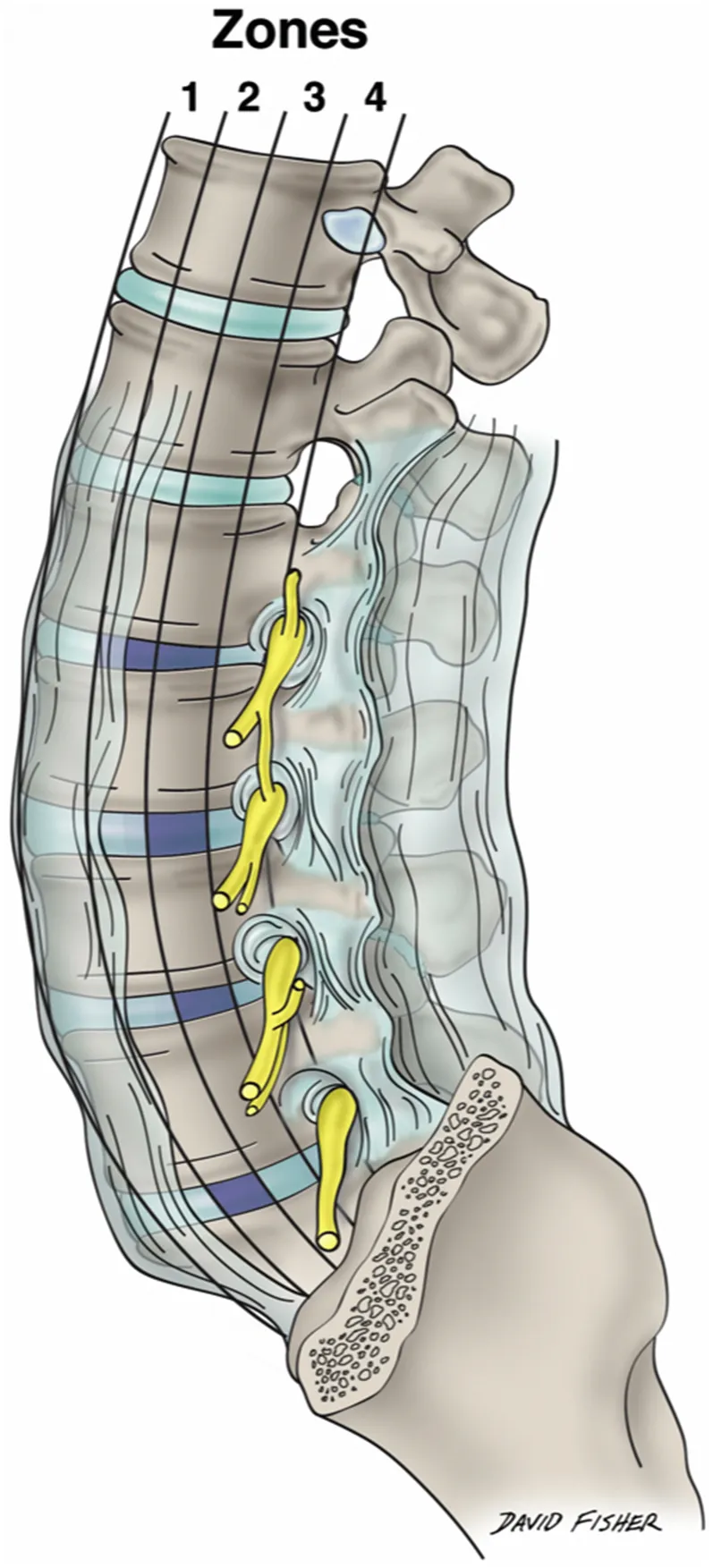
Moro and Uribe et al. subdivided each lumbar vertebral segment into four quarters (zone I to zone IV) from the anterior to the posterior border of the vertebral body (Fig. 1.1) (Uribe et al., 2010; Moro et al., 2003). Although describing the relationship of the plexus nerves to the lateral vertebral body surface helps in determining ideal docking points, it oversimplifies the complex plexus anatomy, as the nerves run in anteroposterior, superolateral, and mediolateral directions. As shown in the study of Grunert et al., injuries can occur throughout the entire trajectory of the lateral transpsoas approach to the lumbar spine. Over 50% of the nerve injuries occurred either at the lateral aspect of the psoas major muscle, within the outer abdominal muscles, or in the subcutaneous tissue of the abdominal wall, predominantly affecting the subcostal, ilioinguinal, iliohypogastric, and lateral femoral cutaneous nerves (Grunert et al., 2017).
As the superficial nerves of the region between the iliac crest and 12th rib are so concentrated (Fig. 1.2) and important to avoid with the lateral transpsoas approach to the lumbar spine, this chapter focuses on these structures and their detailed anatomy.
Segmental Lateral Cutaneous Branches
The skin and muscles of the anterolateral abdominal wall are innervated by the ventral rami of T7–T12. The muscles here also receive fibers from at least L1 ventral ramus and, as we have reported, as inferior as an L4 contribution. The lower thoracic and L1 nerves and their branches travel into the abdominal wall between the transversus abdominis and internal abdominal oblique muscles. Distally, they pierce the rectus sheath. Along their course, the nerves supply not only the skin and adjacent musculature but also the parietal peritoneum. These nerves give rise to lateral and anterior cutaneous branches (Figs. 1.3 and 1.4). The former arise at about the anterior axillary line and pierce the anterolateral muscles of the abdominal wall near the midaxillary line. As the lateral cutaneous branches of these nerves reach the skin, they split into anterior and posterior branches. The anterior cutaneous branches are the terminal branches of each of these segmental nerves and exit the rectus sheath anteriorly to reach the overlying skin where they too split into branches, medial and lateral.
Superior Cluneal Nerves
The superior cluneal nerves (SCNs) (Figs. 1.5–1.8) are the posterior cutaneous branches (from the lateral branch) of the dorsal rami usually described as arising from the upper three lumbar spinal nerves. Historically, it has been believed that the origin of the SCN is the dorsal rami of the L1, L2, and L3 spinal nerves. The SCN is usually depicted as having three branches: medial, intermediate, and lateral SCN.
Out of 20 sides, we previously reported the vertebral level of the origin of the SCN was T12 on 2 sides (10%), L1 on 15 sides (75%), L2 on 18 sides (90%), L3 on 19 sides (95%), L4 on 9 sides (45%) (Fig. 1.9), and L5 on 2 sides (10%), respec...