Following a question-answer approach throughout the narrative, with self-assessment MCQs, EMQs and SAQs, Cardiology: Clinical Cases Uncovered includes sections on cardiac anatomy, physiology and pathology which provide the essentials required to understand clinical cardiology, and is ideal for medical students and junior doctors on the Foundation Programme, specialist nurses and nurse practitioners, and for those with plans for a career in cardiology.
- English
- ePUB (mobile friendly)
- Available on iOS & Android
eBook - ePub
About this book
Cardiology: Clinical Cases Uncovered is the ideal integrated text to help you recognize, understand and know how to investigate and manage many heart-related disorders and conditions. Written by three practising cardiologists, it leads students through a clinical approach to managing problems with 26 real-world cardiovascular cases. There is strong emphasis on high-quality figures, particularly 12-lead ECGs, as these play such a major role in the evaluation of the cardiac patient.
Trusted by 375,005 students
Access to over 1.5 million titles for a fair monthly price.
Study more efficiently using our study tools.
Information
Part 1
Basics
Basic science
Anatomy
The primary function of the heart is to pump deoxygenated blood to the lungs and to return oxygenated blood to the rest of the body. The basic anatomy consists of:
- Pericardium (visceral and parietal): the fibrous sac containing the heart.
- Four cardiac chambers: the right and left atria and ventricles.
- Heart valves:
- Two outflow valves: the aortic and pulmonary valves consist of three semi-lunar cusps.
- Two atrioventricular (AV) valves: the mitral and tricuspid valves, which are attached by chordae tendinae to papillary muscles.
- Vascular system:
- Great vessels: the pulmonary artery, pulmonary vein and aorta.
- Three main coronary arteries: the left anterior descending (LAD) and circumflex (Cx) arteries, which originate from the left main stem (LMS) and the right coronary artery (RCA).
- Venous system: the venous blood is drained via the great cardiac vein, small anterior cardiac vein and thesbian veins.
- Electrical conducting system, which consists of specialised cells that are able to depolarise spontaneously (automaticity) forming:
- The sinoatrial (SA) node.
- The atrioventricular node.
- The Bundle of His (right and left) and terminal Purkinje fibres.
The foetal heart
A knowledge of basic cardiac embryology is helpful for understanding how lesions found in adult congenital heart disease develop.
Foetal atria and ventricles (Figure A )
The heart begins life as a primitive tube, which folds to produce early cardiac chambers: the sinus venosus, the primitive atrium, the ventricle and the bulbus cordis. Further separation of the chambers occurs as follows:
- A pair of septa, the septum primum and septum secundum, grow to separate the right and left atria. The septum primum fuses with the endocardial cushions, the septum secundum does not. The free edge of the septum primum and secundum form the foramen ovale.
- A muscular interventricular septum grows from the floor of the common ventricle to divide it into two chambers.
Foetal shunts (Figure B )
The lungs are bypassed in the foetal circulation by the following right to left shunts:
- Foramen ovale: oxygenated blood passes from the left umbilical vein to the right atrium via the ductus venosus. From the right atrium the blood is then shunted through the foramen ovale to the left atrium
- Ductus arteriosus: the remaining oxygenated blood passes from the right atrium to the right ventricle and enters the pulmonary trunk. From here it passes via the ductus arteriosus directly to the aorta, bypassing the lungs
Circulation changes at birth
As the newborn takes its first breath, the pulmonary vascular resistance drops and conversion from the foetal to adult circulation starts. The following changes occur:
- The foramen ovale closes by the mechanical effect of the reversal in pressure between the two atria, and forms the fossa ovalis in adult life.
- Changes in oxygen concentration of the blood and hormonal changes contribute to the closure of the ductus arteriosus.
Figure A Development of the heart. LA, left atrium; LV, left ventricle; RA, right atrium; RV, right ventricle.
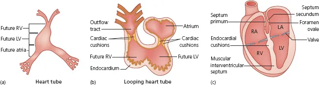
Figure B Foetal circulation and shunts.
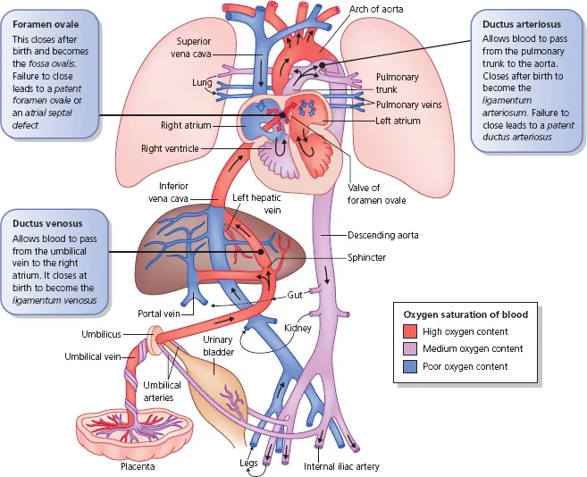
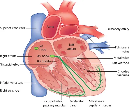
Figure C Adult heart. AV, atrioventricular; SA, sinoatrial.
The adult heart
Right atrium
This chamber is a low-pressure (0–7 mmHg), thin-walled receiving chamber for systemic and cardiac venous blood. It also contains the ‘pacemaker’ (SA node) and the AV node of the heart
Right ventricle
This chamber receives the venous blood from the right atrium and ejects it into the pulmonary artery. Unlike the left ventricle, it is heavily trabeculated. It contains the moderator band, which contains part of the conduction system, and the papillary muscles of the tricuspid valve. The pressure in this chamber is 15–30 mmHg during systole.
Left atrium
This chamber receives oxygenated blood from the pulmonary veins. Clinically important structures are:
- Pulmonary veins: in normal hearts four pulmonary veins (two upper and two lower) drain oxygenated blood from the lungs into the left atrium.
- Left atrial appendage: a blind-ending sac related to the left atrium and a common site for thrombus formation in patients with atrial fibrillation.
- The pressure in this chamber is slightly higher than in the right atrium (4–12 mmHg).
Left ventricle
This is a high-pressure (90–140 mmHg), thick-walled chamber, which reflects its greater contractile performance. It delivers oxygenated blood systemically. It contains the mitral valve papillary muscles. These are conical muscular projections from the walls of the left ventricle that attach to the chordae tendinae to support the two cusps of the mitral valve.
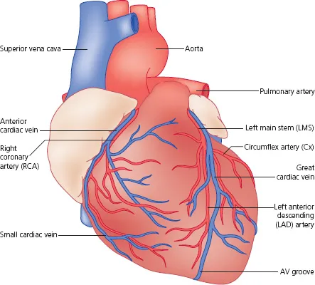
Vascular anatomy (Figure D )
Great vessels
- Superior and inferior vena cava: drain systemic deoxygenated venous blood into the right atrium.
- Pulmonary artery: carries deoxygenated blood to the lungs from the right ventricle. It has thinner walls than systemic arteries and subdivides many times into branches that carry blood to the network of 280 billion capillaries where it is oxygenated.
- Pulmonary veins: there are four draining oxygenated blood from the lungs into the left atrium.
- Aorta: carries oxygenated blood from the left ventricle to supply the rest of the body.
Figure D Vascular anatomy.
Box A Clinical reasons to know cardiac embryology
- A patent foramen ovale (PFO) is found in up to 20% of the population. The majority of people with a PFO have no symptoms. In some patients emboli form in the venous circulation and pass via the patent foramen into the systemic circulation, causing a stroke. (This is known as paradoxical embolus.) In such patients and some other selected groups, closure of the PFO is recommended. This can be done percutaneously.
- Failure of the ductus arteriosus to close after birth leads to the congenital heart defect patent ductus arteriosus (PDA). Surgical or percutaneous duct closure is recommended.
- Failure of the interventricular septum to fuse...
Table of contents
- Cover
- Contents
- Dedication
- Title page
- Copyright page
- Preface
- How to use this book
- List of abbreviations
- Part 1: Basics
- Part 2: Cases
- Part 3: Self-assessment
- Index of cases by diagnosis
- Index
Frequently asked questions
Yes, you can cancel anytime from the Subscription tab in your account settings on the Perlego website. Your subscription will stay active until the end of your current billing period. Learn how to cancel your subscription
No, books cannot be downloaded as external files, such as PDFs, for use outside of Perlego. However, you can download books within the Perlego app for offline reading on mobile or tablet. Learn how to download books offline
Perlego offers two plans: Essential and Complete
- Essential is ideal for learners and professionals who enjoy exploring a wide range of subjects. Access the Essential Library with 800,000+ trusted titles and best-sellers across business, personal growth, and the humanities. Includes unlimited reading time and Standard Read Aloud voice.
- Complete: Perfect for advanced learners and researchers needing full, unrestricted access. Unlock 1.5M+ books across hundreds of subjects, including academic and specialized titles. The Complete Plan also includes advanced features like Premium Read Aloud and Research Assistant.
We are an online textbook subscription service, where you can get access to an entire online library for less than the price of a single book per month. With over 1.5 million books across 990+ topics, we’ve got you covered! Learn about our mission
Look out for the read-aloud symbol on your next book to see if you can listen to it. The read-aloud tool reads text aloud for you, highlighting the text as it is being read. You can pause it, speed it up and slow it down. Learn more about Read Aloud
Yes! You can use the Perlego app on both iOS and Android devices to read anytime, anywhere — even offline. Perfect for commutes or when you’re on the go.
Please note we cannot support devices running on iOS 13 and Android 7 or earlier. Learn more about using the app
Please note we cannot support devices running on iOS 13 and Android 7 or earlier. Learn more about using the app
Yes, you can access Cardiology by Tim Betts,Jeremy Dwight,Sacha Bull in PDF and/or ePUB format, as well as other popular books in Medicine & Cardiology. We have over 1.5 million books available in our catalogue for you to explore.