![]()
CHAPTER 1
Fetal Well-Being and Adaptation at Birth
Key topics
- Placental function
- Fetal homeostasis
- Fetal circulation
- Assessment of fetal well-being
- Screening during pregnancy
- Fetal monitoring during labour
- Fetal compromise
Introduction
The discipline of ‘perinatal medicine’ spans the specialties of fetal medicine and neonatology. The obstetrician must have a thorough knowledge of pregnancy and its effects on the mother and fetus, as well as fetal development and physiology. The neonatologist specializes in the medical care of the infant immediately after birth but must also have a thorough understanding of fetal development and physiology. This chapter reviews fetal assessment and physiology to provide the paediatrician and neonatal nurse with a better understanding of normal perinatal adaptation and the adverse consequences arising from maladaptation.
Placental Function
The placenta is a fetal organ that has three major functions: transport, immunity and metabolism.
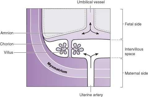
The uterus is supplied with blood from the uterine arteries, which dilate throughout pregnancy, increasing blood supply 10-fold by term. Maternal blood bathes the intervillous space and is separated from fetal blood by the chorionic plate. Transport of nutrients and toxins occurs at this level. Oxygenated fetal blood in the capillaries of the chorionic plate leaves the placenta via the umbilical vein to the fetus (Fig. 1.1).
Transport
The placenta transports nutrients from the mother to the fetus, and waste products in the other direction. This occurs in a number of ways, including simple diffusion (for small molecules) and active transport, which is used for larger molecules. The placenta is crucially also responsible for gaseous exchange of oxygen and carbon dioxide. Oxygen diffuses from the mother (PO2 = 10–14 kPa, 75–105 mmHg) to the fetus (PO2 = 2–4 kPa, 15–30 mmHg) where it binds to fetal haemoglobin. This has a higher affinity for oxygen than maternal haemoglobin for a given PO2. This off-loading of maternal haemoglobin is also facilitated by a change in maternal blood pH.
Immunity
The placenta trophoblast prevents the maternal immune system from reacting against ‘foreign’ fetal antigens. Rejection does not occur because the trophoblastic cells appear to be non-antigenic, although it is known that fetal cells can cross into the maternal circulation where they can trigger an immune reaction (e.g. rhesus haemolytic disease). Maternal IgG antibody, the smallest of the immunoglobulins, can cross the placenta where it provides the newborn with innate immunity to infectious diseases. These IgG antibodies can also cause perinatal disease such as transient hyperthyroidism (see Chapter 21).
CLINICAL TIP: Because IgG antibody crosses the placenta, the presence of IgG antibody in the newborn’s blood does not mean it has been exposed to the disease. This is of particular relevance when testing newborns for HIV infection or syphilis, where a positive IgG may just reflect maternal exposure. Instead, direct tests (e.g. viral RNA by PCR) are required (see Chapter 10).
Metabolism
The placenta is metabolically active and produces hormones, including human chorionic gonadotropin (hCG) and human chorionic thyrotropin (hCT). It also detoxifies drugs and metabolites. Oestriol cannot be produced by the placenta alone. This is done by the fetal liver and adrenal glands. The metabolites are then sulphated by the placenta to form oestrogens, one of which is oestriol.
Because of its metabolic activity, the placenta has very high energy demands and consumes over 50% of the total oxygen and glucose transported across it.
Fetal Homeostasis
The placenta is an essential organ for maintaining fetal homeostasis but the fetus is capable of performing a variety of physiological functions:
- The liver produces albumin, coagulation factors and red blood cells.
- The kidney excretes large volumes of dilute urine from 10–11 weeks’ gestation, which contributes to amniotic fluid.
- Fetal endocrine organs produce thyroid hormones, corticosteroids, mineralocorticoids, parathormone and insulin from 12 weeks’ gestation.
- Some immunoglobulins are produced by the fetus from the end of the first trimester.
Fetal Circulation
The fetal circulation is quite different from the newborn or adult circulation. The umbilical arteries are branches of the internal iliac arteries. These carry deoxygenated blood from the fetus to the placenta where it is oxygenated as it comes into close apposition with maternal blood in the intervillous spaces. Oxygenated fetal blood is carried in the single umbilical vein which bypasses the liver via the ductus venosus to reach the inferior vena cava. It then passes into the inferior vena cava and enters the right atrium as a ‘jet’, which is shunted to the left atrium across the foramen ovale (Fig. 1.2). From here it passes into the left ventricle and is pumped to the coronary arteries and cerebral vessels. In this way the fetal brain receives the most oxygenated blood. Some deoxygenated blood is pumped by the right ventricle into the pulmonary artery, but the majority bypasses the lungs via the ductus arteriosus to flow into the aorta where it is carried back to the placenta. Only 7% of the combined ventricular output of blood passes into the lungs. The right ventricle is the dominant ventricle, ejecting 66% of the combined ventricular output.