![]()
CHAPTER 1
The healthy heart
The human heart – about the size of a clenched fist – is the center of a complex system designed to help the body nourish its organs with life-giving oxygen and to remove waste products in the form of carbon dioxide from the body. Simple animals, such as insects, have an open circulatory system, in which the heart pumps blood through the body cavity, washing the organs directly. More complex animals, including all vertebrates, have a closed circulatory system, which requires the heart to pump blood throughout a network of vessels. No system of vessels is as complex as that of a human being, and it is so closely related to the heart’s function that we frequently talk of the “cardiovascular” or CV system rather than the heart in isolation.
In the human CV system, blood stays in the vessels while oxygen and carbon dioxide are exchanged by diffusion through the vessel walls. So intricate is the human circulatory system that there are actually two complementary networks: a pulmonary circulatory system, designed to get deoxygenated blood to the lungs so it can be “revitalized” with oxygen, and a systemic circulatory system which pumps oxygen-rich blood throughout the body to nourish muscles, tissues, and organs.
At the heart of this elaborate circulatory system is, literally, the heart. At its most basic, the healthy human heart is an efficient and effective pump, beating about 70 times a minute without stopping over the course of a human lifespan. The circulatory system is designed to get oxygen-rich blood where it is needed when it is needed, so the heart regulates its own activities, beating more rapidly during times of increased oxygen consumption and more slowly during periods of decreased demand, such as rest and sleep.
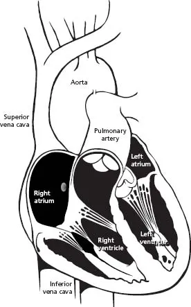
The heart is a muscle with four hollow chambers: two upper and two lower (Fig. 1.1). On top are the atria (singular: atrium), thin-walled, small chambers that take their name from our architectural word “atrium.” They are the ante-chambers or front lobby of the heart. The two lower chambers are large, thick-walled, heavily muscled chambers called ventricles. The ventricles are responsible for most of the pumping action of the heart.
While physicians can talk about the heart in terms of atria and ventricles, or upper and lower chambers, it is also possible to talk about the heart in terms of right side and left side. The right side of the heart consists of the right atrium and the right ventricle, which are connected to each other through the tricuspid valve. When deoxygenated blood flows back to the heart to become reoxygenated, it first enters the right side of the heart. This deoxy-genated blood arrives at the right side of the heart through the body’s largest veins: the superior vena cava and the inferior vena cava. (In this case, “superior” and “inferior” refer to physical locations of “above” and “below” the heart.) The right side of the heart pumps this oxygen-depleted blood back out through the pulmonary artery to the lungs, where it picks up much-needed oxygen. Once the blood has received oxygen, the venous system routes the blood back into the heart, this time to the left side.
Loaded with oxygen, the blood reenters the heart through the pulmonary veins into the left atrium and the left ventricle, connected by the mitral valve. The left side of the heart pumps the blood back out to the rest of the body (the systemic circulatory system) through the aorta and the arteries that branch off the aorta.
Cardiac pacing and defibrillation leads for conventional pacemaker and implantable cardioverter defibrillator (ICD) systems are implanted in the right side of the heart. A transvenous lead – i.e. a wire that goes through a patient’s vein – can “go with the flow” of blood into the right atrium, through the tricuspid valve, and into the right ventricle. Conventional pacemakers and ICDs have found that pacing the right side of the heart is sufficient to cause a contraction of the entire heart. More recent cardiac resynchronization therapy (CRT) devices require a pacing lead to be implanted in both the right and the left sides of the heart. This poses some technical challenges as a transvenous lead cannot readily travel to this area without going through the heart and then against the heart’s natural powerful pumping action. CRT therapy and lead placement falls outside the scope of this book, but it is mentioned to give the reader a more complete view of the therapies available.
Myocardial cells
The heart pumps blood through rhythmic contractions or beats, also known as “depolarizations.” Depolarization explains what happens to the heart at the cellular level, which is the best way to understand how it beats. Unlike other muscles, which respond to the control of the brain, the heart regulates its own actions without specific input from the brain. To accomplish this, it relies on some of the body’s most complex cellular constructions and interactions.
The healthy human heart has two main types of cells: myocardial cells (heart muscle cells) and conduction system cells (electrical cells). Myocardial cells are the ones that make the heart beat.
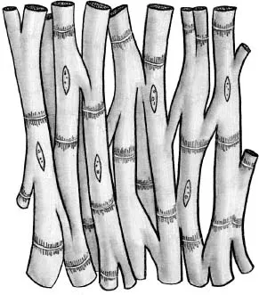
All heart cells are cylindrical and branch at their ends into one or more limbs. These cardiac cells are held together with intercalated disks sandwiched between them to form a network (Fig. 1.2). Think of myocardial cells as a dense forest of trunks and limbs and branches with intercalated disks forming connections. These intercalated disks help conduct electricity from cell to cell by relaying the impulse.
While myocardial cells do not conduct electricity as rapidly as the electrical cells of the heart, they do have the property of contractility, an ability to shorten and then return to their original length. Contractility allows myocardial cells to stretch and snap back into place. In this way, the myocardium or heart muscle is able to expand to take in blood and then to contract powerfully to pump the blood back out.
Table 1.1 Common cardiac drugs
|
| Sympathomimetics (digitalis, bretylium) | X | |
| Beta blockers | | X |
| Quinidine | | X |
| Procainamide | | X |
| Excessive potassium | | X |
| Hypovolemia | X | |
| Anemia | X | |
| Hypocalcemia | | X |
| Hypothyroidism | | X |
| Emotion | X | |
| Increased venous return to the heart | X | |
| Shock | | X |
| Fever | X | |
| Exercise | X | |
| Emotion | X |
Myocardial contractility responds to a variety of influences. Physical stimuli (including exercise, emotion, fever) and some drugs (sympathomimetics such as digitalis) can increase myocardial contractility, forcing the heart to beat more vigorously. Likewise, other stimuli (shock, hypothyroidism, and others) and some drugs (beta blockers, quinidine, procainamide, and excess potassium) can decrease myocardial contractility (Table 1.1).
The heartbeat
An electrical impulse traveling through the heart causes the cardiac cells to depolarize and contract. The human heartbeat is not one single contraction, but is a precisely timed sequence of four specific events (Fig. 1.3).
Starting with the heart at rest, blood flows naturally into the heart. The valves are open and the heart gets a considerable amount of blood into it through a descriptively named process known as the passive filling of the ventricles. The atria are relaxed in a state known as atrial diastole. When an electrical impulse fires in the heart, the heart beat begins its four-part cycle.
The atria contract (atrial systole) while the ventricles remain relaxed (ventricular diastole). Since the ventricles are already passively filled with blood, this atrial contraction forces even more blood into the ventricles. Known as the atrial contribution to ventricle filling (or “atrial kick”) this atrial contraction ensures that the ventricles are filled to the point where they have to stretch to accommodate all of the blood within them. The valves joining atrial to ventricular chambers close, so the ventricles now contain a great deal of blood that cannot backflow into the atria.
There is a brief period of rest – me...