![]()
Part 1
Anatomy and Conformation
![]()
Chapter 1
The whole horse
Introduction (Fig. 1.1)
This book is designed to help all horse owners and riders understand how a horse moves and how its anatomy helps it to perform. It will also help riders recognise the limitations imposed on performance by the horse’s own physical make-up.
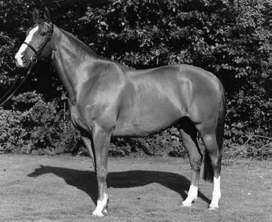
Contrary to popular belief the horse, although a natural athlete, is not a natural jumper. A loose horse, given enough excitement, will naturally perform movements that equate to passage, piaffe, courbette and capriole; however, the horse’s heavy gut and relatively inflexible spine combine to make jumping more difficult. Figure 1.1 shows the template of the whole horse used for illustrative purposes throughout this book.
Fig. 1.1 The whole horse
Training the performance horse
Observation of the following points when exercising and training the performance horse will help to protect bones, tendons, ligaments and muscles from injury:
- Feeding a balanced diet will ensure that the necessary nutrients for energy production, and tissue maintenance and repair are available.
- Water is essential to maintain all body functions; therefore a supply of fresh clean water must be available.
- The salts lost in sweat should be replaced in the form of electrolytes in the diet; these body salts are vital for nerve and muscle function.
- Thorough warming up before executing strenuous or difficult exercises will ensure adequate blood supply to the muscles and minimise the rise of injury.
- Thorough cooling down after exercise will help the circulation remove the toxic waste products of exercise (for example, lactic acid) and reduce muscle stiffness.
- Allowing the horse periods of relaxation and stretching during work will help to prevent tension. When muscles are held in tension (think how your shoulders feel after a stressful drive) the blood supply is inadequate to remove waste products which accumulate and increase the risk of injury and stiffness.
- Regular shoeing to keep the horse’s feet in balance will ensure that the joints of the limb remain in correct alignment and that the forces generated during work are transmitted up the limb in a way that minmises the risk of injury.
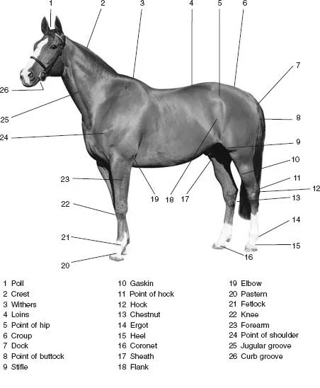
Points of the horse (Fig. 1.2)
Familiarity with the surface anatomy of the horse is important so that underlying structures can be identified. The horse has evolved over many thousands of years from a small fox-like creature with four toes on each foot to the animal it is today with a single hoof at the end of each limb. Figure 1.2 shows the points of the horse in detail.
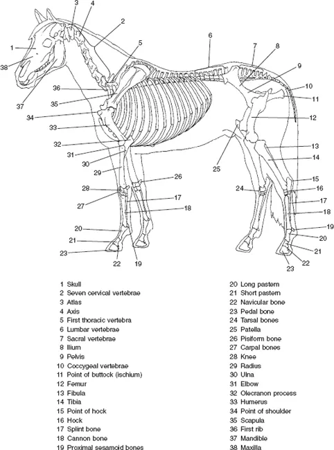
The skeleton (Fig. 1.3)
The skeleton is the framework that supports and protects the soft tissues of the horse’s body. In Fig. 1.2 many bony areas lying below the skin are identified. Figure 1.3 shows the positions of the bones that give rise to some of the points of the horse.
Fig. 1.2 Points of the horse
Functions of the skeleton
- The skeleton is made of bone.
- Bone protects soft tissue; for example, the skull encloses and protects the brain, and the vertebral column encloses and protects the spinal cord.
- Skeletal muscles are attached to bones via tendons to enable movement.
- Bone is a living tissue with a supply of blood and nerves.
- Bones meet and articulate at joints that vary in their ability to move.
- The ends of the bones making up movable joints are covered by cartilage, allowing them to slide smoothly over one another.
- Ligaments bind bone to bone, supporting the joints.
- The interior of the joint is lubricated by synovial fluid which is secreted by the membrane covering the cartilage at the ends of the bones.
Fig. 1.3 Skeleton
Skeletal muscles
The horse’s skeleton is incapable of movement on its own. All movements, from a flick of the tail to the most difficult dressage manoeuvre, are brought about by a complicated system of skeletal muscles. All horses, regardless of breed, size or age, have the same arrangement of skeletal muscles, but some muscles may be better developed in certain horses depending upon their type of training. For example, the Thoroughbred dressage horse will have a more highly developed topline than a racing-fit Thoroughbred. In addition, centuries of selective breeding have led to enhanced muscular development in some breeds and types of horses. The Quarter horse, bred to sprint over a quarter of a mile, has highly muscular forelimbs and hindquarters. The sprinting Thoroughbred naturally has a more muscular physique than its steeplechasing counterpart.
The skeletal muscles are under the horse’s conscious control and enable it to adjust to the surrounding environment and to make necessary movements such as running or grazing. Muscles are attached to and hence move, various parts of the skeleton and body.
Skeletal muscles create movement by acting across joints. They are usually arranged in opposing groups which perform opposite actions to give smooth and even movements. Flexors are placed behind the bone and pull it backwards, i.e. bend the joint, whereas extensors are placed in front of the bone and pull it forwards, i.e. straighten the joint from the bent position. Usually one of the pair of muscles is much stronger than the other.
Each end of the muscle tapers from a larger muscle belly into a tendon, which attaches the muscle to the bone. Muscle bellies vary in size and shape: some are large flat sheets, such as the latissimus dorsi, and others are long and strap-like, such as the brachiocephalicus.
The horse has no muscles below the knee, and all movement in this area is carried out via tendons attached to the muscles higher up the limb. This results in the lower limbs of performance horses being prone to tendon and ligament injury. Tired muscles are more likely to result in injury.
For muscles to produce movement, they must be attached to bone at both ends. These ends are sometimes classified as the ‘origin’ and ‘insertion’, the origi...