- 224 pages
- English
- ePUB (mobile friendly)
- Available on iOS & Android
eBook - ePub
The Auditory System in Sleep
About this book
The Auditory System in Sleep presents for the first time a view of a sensory system working in a different state-that of the sleeping brain. The auditory system is always "open receiving information from the environment and the body itself (conscious and unconscious data). Even during sleep the auditory information is processed, although in a different way. This book draws information from evoked potentials, fMRI, PET, SPECT, lesions, etc., together with electrophysiological online data in order to depict how the auditory system single unit activity, recorded during sleep, revealed the possibility of sensory information participation in sleep processes.
- Presents diverse experimental viewpoints from the beginning of classical electroencephalography to the more recent imaging, single units, electro-magneto-encephalography studies, etc.
- Includes classic data as well as new data based in the existing literature and on the long scientific research lines (auditory and sleep) developed by the author and coworkers on this subject since 1963
Tools to learn more effectively

Saving Books

Keyword Search

Annotating Text

Listen to it instead
Information
CHAPTER 1
Brief analysis of the organization of the auditory system and its physiological basis
Publisher Summary
This chapter gives the review of the known auditory ascending and descending systems that is presented along with some new or not well-known approaches. The auditory system with its associated anatomical and functional complexity serves diverse processes such as discrimination of sound frequencies and intensities, sound source location in space, auditory learning, development of human language, auditory “images” in dreams, music, development of birds’ songs, i.e., communication in general. The afferent ascending system is a complex system, which begins at the receptors in the cochlea followed by a wide upward expansion throughout the different nuclei, reticular formation, cerebellum, and connections to the primary and secondary cortices. It is composed of several neuronal groups with profuse communication from the cochlea to the cortex. A diagram of the most important pathways and synaptic stations of the afferent auditory system is provided in this chapter. The auditory pathway has been described by using different methods of study throughout history: cell damage and degeneration, intracellular dyeing with tracers, deoxyglucose, and so on, and also by electrophysiological recording methods. This study briefly describes the auditory nerve evoked activity; the auditory nerve compound action potential (cAP) can be recorded from an electrode placed at the round window. Along with this, superior olivary complex, inferior colliculus, medial geniculate body and many more are briefly discussed in this chapter.
The auditory system with its associated anatomical and functional complexity subserves diverse processes such as discrimination of sound frequencies and intensities, sound source location in space, auditory learning, development of human language, auditory “images” in dreams, music, development of birds songs, i.e., communication in general. In this chapter a review of the known auditory ascending and descending systems is presented along with some new or not well-known approaches included.
The afferent ascending system
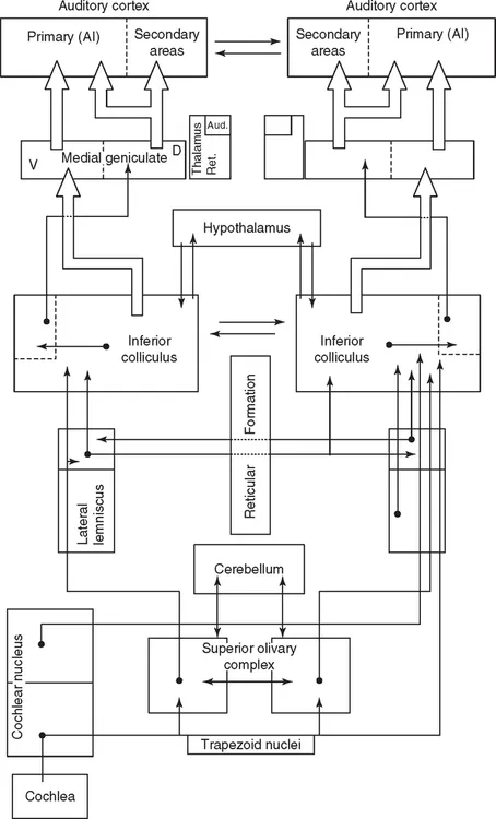
This complex system begins at the receptors in the cochlea followed by a wide upward expansion throughout the different nuclei, reticular formation, cerebellum, and connections to the primary and secondary cortices. It is composed of several neuronal groups with profuse communication from the cochlea to the cortex. A non-classical ascending pathway branches off from classical IC and reaches the medial geniculate nucleus, medial and dorsal regions, to project to cortical regions (Moller and Rollins, 2002).
A diagram of the most important pathways and synaptic stations of the afferent auditory system is shown in Figure 1.1. The first-order auditory neurones, with cell bodies located in Corti’s ganglion, send their axons centrally to form the auditory nerve, part of the VIIIth cranial pair. These nerve fibers synapse with the secondary neurones located centrally in different cochlear nucleus (CN) loci, in the medulla–pontine region. Let us bear in mind that 95% of the fibers which form the auditory nerve originate at the inner hair cells. The outer hair cells are innervated by only 5%, non-myelinated afferent thin fibers.
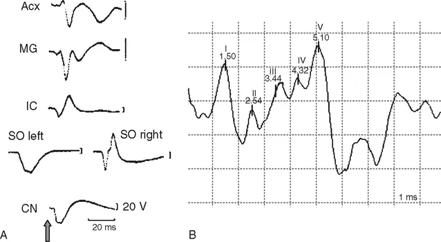
The auditory pathway has been described by using different methods of study throughout history: cell damage and degeneration, intracellular dyeing with tracers, deoxyglucose, and so on, and also by electrophysiological recording methods. By placing recording electrodes in various central nuclei, bioelectrical responses – changes in the membrane potentials – can be obtained of the auditory neurones which form the basis of evoked potentials measurable with gross electrode. Evoked potentials, recorded in cats, shown in Figure 1.3(A), are examples of the averaged responses to brief (click) sound stimuli. The differences between their shapes and, mainly, their latencies carefully reproduce the anatomical pathway, due to the fact that activity evoked by a stimulus first activate the receptors, followed by the auditory nerve fibers, and subsequently the central nervous system (CNS), orderly ascending from nucleus to nucleus.
Auditory nerve evoked activity
Beginning with the incoming sound, Table 1.1 exhibits the main mechanobioelectrical steps toward evoking an auditory nerve action potential. The auditory nerve compound action potential (cAP) can be recorded from an electrode placed at the round window. A cAP averaged is depicted in Figure 1.2 (left) with the two classical negative waves N1 and N2, in response to clicks, i.e., a stimulus that synchronizes the discharge of many nerve fibers. It reveals the activity of a group of single fibers and its synchronized discharges. The N1 amplitude is a function of the stimulus intensity as well as the number of synchronized fibers.
TABLE 1.1
Events toward the generation of an auditory nerve action potential
Sound waves move the tympanic membrane
↓
The tympanic membrane moves the middle-ear ossicles
↓
The ossicles move the oval window membrane
↓
The oval window movements produce motion of the cochlear fluids and basilar membrane
↓
The cochlear fluids and basilar membrane motion bend the inner hair cells’ cilia
↓
The ciliary movements determine the excitation of the hair cells
↓
Finally, action potentials are generated at the auditory nerve fibers
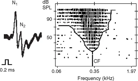
An auditory nerve single-fiber recording is shown in Figure 1.2 (right). A microelectrode may record the single-fiber activity when a stimulus of sufficient intensity is delivered. Its response can be characterized by a point of maximum sensitivity, i.e., the response at the stimulus frequency with the lowest intensity, the characteris...
Table of contents
- Cover image
- Title page
- Table of Contents
- Foreword
- Acknowledgements
- Introduction
- Chapter 1: Brief analysis of the organization of the auditory system and its physiological basis
- Chapter 2: The physiological bases of sleep
- Chapter 3: Notes on information processing
- Chapter 4: Auditory information processing during sleep
- Chapter 5: Auditory unit activity in sleep
- Chapter 6: Auditory influences on sleep
- Conclusions
- References
- Index
Frequently asked questions
Yes, you can cancel anytime from the Subscription tab in your account settings on the Perlego website. Your subscription will stay active until the end of your current billing period. Learn how to cancel your subscription
No, books cannot be downloaded as external files, such as PDFs, for use outside of Perlego. However, you can download books within the Perlego app for offline reading on mobile or tablet. Learn how to download books offline
Perlego offers two plans: Essential and Complete
- Essential is ideal for learners and professionals who enjoy exploring a wide range of subjects. Access the Essential Library with 800,000+ trusted titles and best-sellers across business, personal growth, and the humanities. Includes unlimited reading time and Standard Read Aloud voice.
- Complete: Perfect for advanced learners and researchers needing full, unrestricted access. Unlock 1.4M+ books across hundreds of subjects, including academic and specialized titles. The Complete Plan also includes advanced features like Premium Read Aloud and Research Assistant.
We are an online textbook subscription service, where you can get access to an entire online library for less than the price of a single book per month. With over 1 million books across 990+ topics, we’ve got you covered! Learn about our mission
Look out for the read-aloud symbol on your next book to see if you can listen to it. The read-aloud tool reads text aloud for you, highlighting the text as it is being read. You can pause it, speed it up and slow it down. Learn more about Read Aloud
Yes! You can use the Perlego app on both iOS and Android devices to read anytime, anywhere — even offline. Perfect for commutes or when you’re on the go.
Please note we cannot support devices running on iOS 13 and Android 7 or earlier. Learn more about using the app
Please note we cannot support devices running on iOS 13 and Android 7 or earlier. Learn more about using the app
Yes, you can access The Auditory System in Sleep by Ricardo Velluti in PDF and/or ePUB format, as well as other popular books in Biological Sciences & Physiology. We have over one million books available in our catalogue for you to explore.