
eBook - ePub
The Auditory System in Sleep
Ricardo Velluti
This is a test
Partager le livre
- 224 pages
- English
- ePUB (adapté aux mobiles)
- Disponible sur iOS et Android
eBook - ePub
The Auditory System in Sleep
Ricardo Velluti
Détails du livre
Aperçu du livre
Table des matières
Citations
À propos de ce livre
The Auditory System in Sleep presents for the first time a view of a sensory system working in a different state-that of the sleeping brain. The auditory system is always "open" receiving information from the environment and the body itself (conscious and unconscious data). Even during sleep the auditory information is processed, although in a different way. This book draws information from evoked potentials, fMRI, PET, SPECT, lesions, etc., together with electrophysiological online data in order to depict how the auditory system single unit activity, recorded during sleep, revealed the possibility of sensory information participation in sleep processes.
- Presents diverse experimental viewpoints from the beginning of classical electroencephalography to the more recent imaging, single units, electro-magneto-encephalography studies, etc.
- Includes classic data as well as new data based in the existing literature and on the long scientific research lines (auditory and sleep) developed by the author and coworkers on this subject since 1963
Foire aux questions
Comment puis-je résilier mon abonnement ?
Il vous suffit de vous rendre dans la section compte dans paramètres et de cliquer sur « Résilier l’abonnement ». C’est aussi simple que cela ! Une fois que vous aurez résilié votre abonnement, il restera actif pour le reste de la période pour laquelle vous avez payé. Découvrez-en plus ici.
Puis-je / comment puis-je télécharger des livres ?
Pour le moment, tous nos livres en format ePub adaptés aux mobiles peuvent être téléchargés via l’application. La plupart de nos PDF sont également disponibles en téléchargement et les autres seront téléchargeables très prochainement. Découvrez-en plus ici.
Quelle est la différence entre les formules tarifaires ?
Les deux abonnements vous donnent un accès complet à la bibliothèque et à toutes les fonctionnalités de Perlego. Les seules différences sont les tarifs ainsi que la période d’abonnement : avec l’abonnement annuel, vous économiserez environ 30 % par rapport à 12 mois d’abonnement mensuel.
Qu’est-ce que Perlego ?
Nous sommes un service d’abonnement à des ouvrages universitaires en ligne, où vous pouvez accéder à toute une bibliothèque pour un prix inférieur à celui d’un seul livre par mois. Avec plus d’un million de livres sur plus de 1 000 sujets, nous avons ce qu’il vous faut ! Découvrez-en plus ici.
Prenez-vous en charge la synthèse vocale ?
Recherchez le symbole Écouter sur votre prochain livre pour voir si vous pouvez l’écouter. L’outil Écouter lit le texte à haute voix pour vous, en surlignant le passage qui est en cours de lecture. Vous pouvez le mettre sur pause, l’accélérer ou le ralentir. Découvrez-en plus ici.
Est-ce que The Auditory System in Sleep est un PDF/ePUB en ligne ?
Oui, vous pouvez accéder à The Auditory System in Sleep par Ricardo Velluti en format PDF et/ou ePUB ainsi qu’à d’autres livres populaires dans Ciencias biológicas et Neurociencia. Nous disposons de plus d’un million d’ouvrages à découvrir dans notre catalogue.
Informations
Sujet
Ciencias biológicasSous-sujet
NeurocienciaCHAPTER 1
Brief analysis of the organization of the auditory system and its physiological basis
Publisher Summary
This chapter gives the review of the known auditory ascending and descending systems that is presented along with some new or not well-known approaches. The auditory system with its associated anatomical and functional complexity serves diverse processes such as discrimination of sound frequencies and intensities, sound source location in space, auditory learning, development of human language, auditory “images” in dreams, music, development of birds’ songs, i.e., communication in general. The afferent ascending system is a complex system, which begins at the receptors in the cochlea followed by a wide upward expansion throughout the different nuclei, reticular formation, cerebellum, and connections to the primary and secondary cortices. It is composed of several neuronal groups with profuse communication from the cochlea to the cortex. A diagram of the most important pathways and synaptic stations of the afferent auditory system is provided in this chapter. The auditory pathway has been described by using different methods of study throughout history: cell damage and degeneration, intracellular dyeing with tracers, deoxyglucose, and so on, and also by electrophysiological recording methods. This study briefly describes the auditory nerve evoked activity; the auditory nerve compound action potential (cAP) can be recorded from an electrode placed at the round window. Along with this, superior olivary complex, inferior colliculus, medial geniculate body and many more are briefly discussed in this chapter.
The auditory system with its associated anatomical and functional complexity subserves diverse processes such as discrimination of sound frequencies and intensities, sound source location in space, auditory learning, development of human language, auditory “images” in dreams, music, development of birds songs, i.e., communication in general. In this chapter a review of the known auditory ascending and descending systems is presented along with some new or not well-known approaches included.
The afferent ascending system
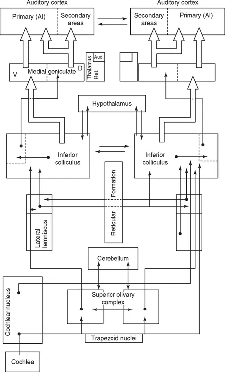
This complex system begins at the receptors in the cochlea followed by a wide upward expansion throughout the different nuclei, reticular formation, cerebellum, and connections to the primary and secondary cortices. It is composed of several neuronal groups with profuse communication from the cochlea to the cortex. A non-classical ascending pathway branches off from classical IC and reaches the medial geniculate nucleus, medial and dorsal regions, to project to cortical regions (Moller and Rollins, 2002).
A diagram of the most important pathways and synaptic stations of the afferent auditory system is shown in Figure 1.1. The first-order auditory neurones, with cell bodies located in Corti’s ganglion, send their axons centrally to form the auditory nerve, part of the VIIIth cranial pair. These nerve fibers synapse with the secondary neurones located centrally in different cochlear nucleus (CN) loci, in the medulla–pontine region. Let us bear in mind that 95% of the fibers which form the auditory nerve originate at the inner hair cells. The outer hair cells are innervated by only 5%, non-myelinated afferent thin fibers.
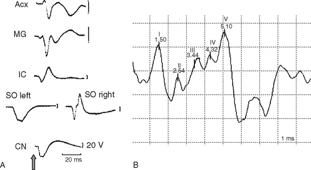
The auditory pathway has been described by using different methods of study throughout history: cell damage and degeneration, intracellular dyeing with tracers, deoxyglucose, and so on, and also by electrophysiological recording methods. By placing recording electrodes in various central nuclei, bioelectrical responses – changes in the membrane potentials – can be obtained of the auditory neurones which form the basis of evoked potentials measurable with gross electrode. Evoked potentials, recorded in cats, shown in Figure 1.3(A), are examples of the averaged responses to brief (click) sound stimuli. The differences between their shapes and, mainly, their latencies carefully reproduce the anatomical pathway, due to the fact that activity evoked by a stimulus first activate the receptors, followed by the auditory nerve fibers, and subsequently the central nervous system (CNS), orderly ascending from nucleus to nucleus.
Auditory nerve evoked activity
Beginning with the incoming sound, Table 1.1 exhibits the main mechanobioelectrical steps toward evoking an auditory nerve action potential. The auditory nerve compound action potential (cAP) can be recorded from an electrode placed at the round window. A cAP averaged is depicted in Figure 1.2 (left) with the two classical negative waves N1 and N2, in response to clicks, i.e., a stimulus that synchronizes the discharge of many nerve fibers. It reveals the activity of a group of single fibers and its synchronized discharges. The N1 amplitude is a function of the stimulus intensity as well as the number of synchronized fibers.
TABLE 1.1
Events toward the generation of an auditory nerve action potential
Sound waves move the tympanic membrane
↓
The tympanic membrane moves the middle-ear ossicles
↓
The ossicles move the oval window membrane
↓
The oval window movements produce motion of the cochlear fluids and basilar membrane
↓
The cochlear fluids and basilar membrane motion bend the inner hair cells’ cilia
↓
The ciliary movements determine the excitation of the hair cells
↓
Finally, action potentials are generated at the auditory nerve fibers
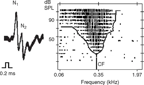
An auditory nerve single-fiber recording is shown in Figure 1.2 (right). A microelectrode may record the single-fiber activity when a stimulus of sufficient intensity is delivered. Its response can be characterized by a point of maximum sensitivity, i.e., the response at the stimulus frequency with the lowest intensity, the characteris...